中国组织工程研究 ›› 2019, Vol. 23 ›› Issue (23): 3705-3709.doi: 10.3969/j.issn.2095-4344.1317
• 组织构建细胞学实验 cytology experiments in tissue construction • 上一篇 下一篇
肌成纤维细胞在小鼠肝内胆管发育过程中的作用
杨均均1,李德卫2,王 伟2,冯 渊1
- (1川北医学院第二临床学院•南充市中心医院感染科,四川省南充市 637000;2重庆医科大学附属第一医院肝胆外科,重庆市 400016)
Role of myofibroblasts in the development of mouse intrahepatic bile ducts
Yang Junjun1, Li Dewei2, Wang Wei2, Feng Yuan1
- (1Department of Infections, the Second Clinical College of North Sichuan Medical College, Nanchong Central Hospital, Nanchong 637000, Sichuan Province, China; 2Department of Hepatobiliary Surgery, the First Affiliated Hospital of Chongqing Medical University, Chongqing 400016, China)
摘要:
文章快速阅读:
.jpg) 文题释义:
细胞角蛋白19(cytokeratin19,CK19):通常作为胆管细胞的标志,在人类肝祖细胞中低表达,但在鼠类肝祖细胞中不表达,在胆管细胞分化早期检测细胞角蛋白19存在技术上的困难。
肌成纤维细胞:是指含有肌动蛋白、肌球蛋白和其他肌肉蛋白的成纤维样细胞。收缩蛋白质排列成可具有收缩功能的形式。成纤维细胞及其表达的细胞因子是肿瘤组织重要的微环境成分,在促进肿瘤的增殖、转移和新生血管形成方面起着非常重要的作用。
文题释义:
细胞角蛋白19(cytokeratin19,CK19):通常作为胆管细胞的标志,在人类肝祖细胞中低表达,但在鼠类肝祖细胞中不表达,在胆管细胞分化早期检测细胞角蛋白19存在技术上的困难。
肌成纤维细胞:是指含有肌动蛋白、肌球蛋白和其他肌肉蛋白的成纤维样细胞。收缩蛋白质排列成可具有收缩功能的形式。成纤维细胞及其表达的细胞因子是肿瘤组织重要的微环境成分,在促进肿瘤的增殖、转移和新生血管形成方面起着非常重要的作用。
文题释义:
细胞角蛋白19(cytokeratin19,CK19):通常作为胆管细胞的标志,在人类肝祖细胞中低表达,但在鼠类肝祖细胞中不表达,在胆管细胞分化早期检测细胞角蛋白19存在技术上的困难。
肌成纤维细胞:是指含有肌动蛋白、肌球蛋白和其他肌肉蛋白的成纤维样细胞。收缩蛋白质排列成可具有收缩功能的形式。成纤维细胞及其表达的细胞因子是肿瘤组织重要的微环境成分,在促进肿瘤的增殖、转移和新生血管形成方面起着非常重要的作用。摘要
背景:研究表明肌成纤维细胞具有促进肝祖细胞分化为胆管细胞作用,但对于肌成纤维细胞在体内胆管发育中作用仍不清楚。
目的:观察小鼠肝内胆管发育的特点,以及肌成纤维细胞在胆管发育过程中的作用。
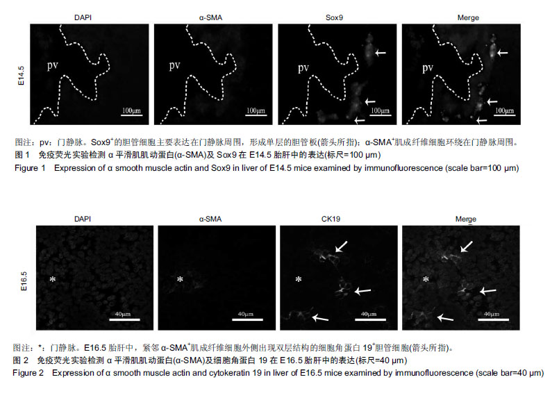
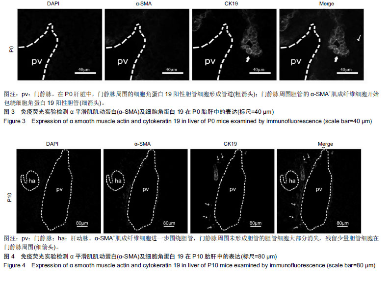
方法:SPF级C57小鼠购自重庆医科大学实验动物中心,实验方案经重庆医科大学动物实验伦理委员会批准,批准号为2017-182。采用14.5 d、16.5 d孕鼠及刚出生(0 d)和出生后10 d的小鼠,麻醉后取其胎鼠肝脏(E14.5、E16.5)和出生后小鼠肝脏(P0、P10),采用免疫组织化学技术观察α平滑肌肌动蛋白、Sox9及细胞角蛋白19在E14.5、E16.5小鼠胎肝和P0、P10小鼠肝脏中的表达。
结果与结论:①E14.5小鼠胎肝中,α平滑肌肌动蛋白阳性肌成纤维细胞环绕门静脉周围,Sox9阳性胆管细胞紧靠α平滑肌肌动蛋白阳性肌成纤维细胞,并形成单层细胞排列的胆管板;②E16.5小鼠胎肝中,紧靠α平滑肌肌动蛋白阳性肌成纤维细胞的细胞角蛋白19阳性胆管细胞形成双层胆管细胞结构;③P0小鼠肝脏中,细胞角蛋白19阳性胆管细胞发育形成具有管腔的胆管结构,并且α平滑肌肌动蛋白阳性肌成纤维细胞开始包绕胆管;④P10小鼠肝脏中,胆管进一步发育扩张,门静脉周围未形成胆管的胆管细胞开始消失。并且α平滑肌肌动蛋白阳性肌成纤维细胞环绕胆管,胆管旁发育形成肝动脉;⑤结果表明了小鼠肝内胆管发育的特点,发现α平滑肌肌动蛋白阳性肌成纤维细胞与肝内胆管发育的关系密切,且在肝内胆管发育中发挥了重要作用。
中国组织工程研究杂志出版内容重点:组织构建;骨细胞;软骨细胞;细胞培养;成纤维细胞;血管内皮细胞;骨质疏松;组织工程 ORCID: 0000-0002-5282-0129(杨均均)
中图分类号:
.jpg) 文题释义:
细胞角蛋白19(cytokeratin19,CK19):通常作为胆管细胞的标志,在人类肝祖细胞中低表达,但在鼠类肝祖细胞中不表达,在胆管细胞分化早期检测细胞角蛋白19存在技术上的困难。
肌成纤维细胞:是指含有肌动蛋白、肌球蛋白和其他肌肉蛋白的成纤维样细胞。收缩蛋白质排列成可具有收缩功能的形式。成纤维细胞及其表达的细胞因子是肿瘤组织重要的微环境成分,在促进肿瘤的增殖、转移和新生血管形成方面起着非常重要的作用。
文题释义:
细胞角蛋白19(cytokeratin19,CK19):通常作为胆管细胞的标志,在人类肝祖细胞中低表达,但在鼠类肝祖细胞中不表达,在胆管细胞分化早期检测细胞角蛋白19存在技术上的困难。
肌成纤维细胞:是指含有肌动蛋白、肌球蛋白和其他肌肉蛋白的成纤维样细胞。收缩蛋白质排列成可具有收缩功能的形式。成纤维细胞及其表达的细胞因子是肿瘤组织重要的微环境成分,在促进肿瘤的增殖、转移和新生血管形成方面起着非常重要的作用。