中国组织工程研究 ›› 2019, Vol. 23 ›› Issue (19): 3050-3055.doi: 10.3969/j.issn.2095-4344.1250
• 组织构建实验造模 experimental modeling in tissue construction • 上一篇 下一篇
甲基强的松龙抑制脊髓损伤模型大鼠小胶质细胞活化介导的炎症
黄星球,廖绪强,史成龙,张秀丽
- (佛山市第一人民医院,广东省佛山市 528000)
Methylprednisolone inhibits inflammation mediated by activated microglia after spinal cord injury in rat models
Huang Xingqiu, Liao Xuqiang, Shi Chenglong, Zhang Xiuli
- (the First People’s Hospital of Foshan, Foshan 528000, Guangdong Province, China)
摘要:
文章快速阅读:
.jpg) 文题释义:
小胶质细胞:系神经组织中唯一来源于中胚层的细胞,其细胞体很小,呈短棒状,伸出数支枯条样突起,突起表面粗糙,显有棘刺,分支少;胞核较小,约为5 μm,形态不规则,可呈肾形、椭圆形或三角形,核染色质多。作为中枢神经系统固有免疫细胞,小胶质细胞被认为是神经炎症的发动者。
糖皮质激素:是由肾上腺皮质中束状带分泌的一类类固醇激素,主要为皮质醇,具有调节糖、脂肪和蛋白质的生物合成和代谢的作用,还具有抑制免疫应答、抗炎、抗毒、抗休克作用。称其为“糖皮质激素”是因为其调节糖类代谢的活性最早为人们所认识,在临床工作中激素因具有较强的抗炎抗变态反应作用而广泛应用于支气管哮喘治疗中。
文题释义:
小胶质细胞:系神经组织中唯一来源于中胚层的细胞,其细胞体很小,呈短棒状,伸出数支枯条样突起,突起表面粗糙,显有棘刺,分支少;胞核较小,约为5 μm,形态不规则,可呈肾形、椭圆形或三角形,核染色质多。作为中枢神经系统固有免疫细胞,小胶质细胞被认为是神经炎症的发动者。
糖皮质激素:是由肾上腺皮质中束状带分泌的一类类固醇激素,主要为皮质醇,具有调节糖、脂肪和蛋白质的生物合成和代谢的作用,还具有抑制免疫应答、抗炎、抗毒、抗休克作用。称其为“糖皮质激素”是因为其调节糖类代谢的活性最早为人们所认识,在临床工作中激素因具有较强的抗炎抗变态反应作用而广泛应用于支气管哮喘治疗中。
文题释义:
小胶质细胞:系神经组织中唯一来源于中胚层的细胞,其细胞体很小,呈短棒状,伸出数支枯条样突起,突起表面粗糙,显有棘刺,分支少;胞核较小,约为5 μm,形态不规则,可呈肾形、椭圆形或三角形,核染色质多。作为中枢神经系统固有免疫细胞,小胶质细胞被认为是神经炎症的发动者。
糖皮质激素:是由肾上腺皮质中束状带分泌的一类类固醇激素,主要为皮质醇,具有调节糖、脂肪和蛋白质的生物合成和代谢的作用,还具有抑制免疫应答、抗炎、抗毒、抗休克作用。称其为“糖皮质激素”是因为其调节糖类代谢的活性最早为人们所认识,在临床工作中激素因具有较强的抗炎抗变态反应作用而广泛应用于支气管哮喘治疗中。摘要
背景:小胶质细胞作为中枢神经系统的固有免疫细胞,已被证实参与调控中枢神经系统损伤后的神经炎症。甲基强的松龙冲击疗法作为目前脊髓损伤早期临床常用治疗方法,其对脊髓损伤后神经炎症的作用及机制尚不清楚。
目的:观察甲基强的松龙对脊髓损伤后小胶质细胞活化介导炎症的作用。
方法:36只SD大鼠购自上海斯莱克实验动物责任有限公司。将大鼠随机分为假手术组、模型组、甲强龙组,每组12只。假手术组仅行椎板切除术;模型组和甲强龙组均采用NYU脊髓打击器制备脊髓损伤模型,甲强龙组给予甲基强的松龙尾静脉注射;假手术组和模型组给予等量0.9%氯化钠尾静脉注射。干预后3 d取材,采用尼氏染色光镜观察形态,Elisa检测大鼠静脉血中白细胞介素1β和白细胞介素18水平,激光共聚焦扫描显微镜观察活化的小胶质细胞IBA-1/ED-1双阳性细胞数,Western blot检测ED-1蛋白表达情况,qPCR检测白细胞介素1β mRNA和白细胞介素18 mRNA表达量。
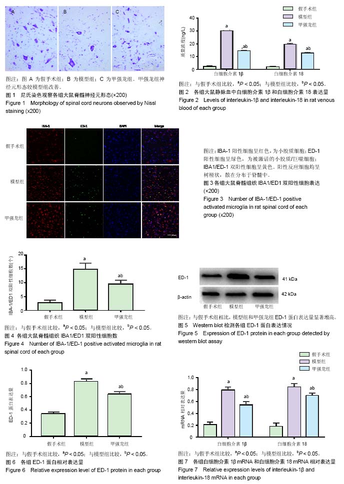
结果与结论:①尼氏染色提示甲基强的松龙可改善脊髓损伤后神经元形态,促进神经元恢复;②与模型组比较,甲强龙组白细胞介素1β和白细胞介素18表达水平显著降低(P < 0.05);③与模型组比较,甲强龙组IBA-1/ED-1双阳性细胞数显著减少(P < 0.05);④与模型组比较,甲强龙组ED-1蛋白表达量显著减少(P < 0.05);⑤与模型组比较,甲强龙组白细胞介素1β mRNA和白细胞介素18 mRNA表达水平显著降低;⑥结果说明,甲基强的松龙抑制炎症的机制与其抑制脊髓损伤后小胶质细胞活化介导的炎症有关,从而有利于神经元恢复。
中国组织工程研究杂志出版内容重点:组织构建;骨细胞;软骨细胞;细胞培养;成纤维细胞;血管内皮细胞;骨质疏松;组织工程
ORCID: 0000-0002-9372-685X(黄星球)
中图分类号:
.jpg) 文题释义:
小胶质细胞:系神经组织中唯一来源于中胚层的细胞,其细胞体很小,呈短棒状,伸出数支枯条样突起,突起表面粗糙,显有棘刺,分支少;胞核较小,约为5 μm,形态不规则,可呈肾形、椭圆形或三角形,核染色质多。作为中枢神经系统固有免疫细胞,小胶质细胞被认为是神经炎症的发动者。
糖皮质激素:是由肾上腺皮质中束状带分泌的一类类固醇激素,主要为皮质醇,具有调节糖、脂肪和蛋白质的生物合成和代谢的作用,还具有抑制免疫应答、抗炎、抗毒、抗休克作用。称其为“糖皮质激素”是因为其调节糖类代谢的活性最早为人们所认识,在临床工作中激素因具有较强的抗炎抗变态反应作用而广泛应用于支气管哮喘治疗中。
文题释义:
小胶质细胞:系神经组织中唯一来源于中胚层的细胞,其细胞体很小,呈短棒状,伸出数支枯条样突起,突起表面粗糙,显有棘刺,分支少;胞核较小,约为5 μm,形态不规则,可呈肾形、椭圆形或三角形,核染色质多。作为中枢神经系统固有免疫细胞,小胶质细胞被认为是神经炎症的发动者。
糖皮质激素:是由肾上腺皮质中束状带分泌的一类类固醇激素,主要为皮质醇,具有调节糖、脂肪和蛋白质的生物合成和代谢的作用,还具有抑制免疫应答、抗炎、抗毒、抗休克作用。称其为“糖皮质激素”是因为其调节糖类代谢的活性最早为人们所认识,在临床工作中激素因具有较强的抗炎抗变态反应作用而广泛应用于支气管哮喘治疗中。