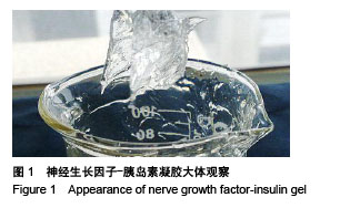
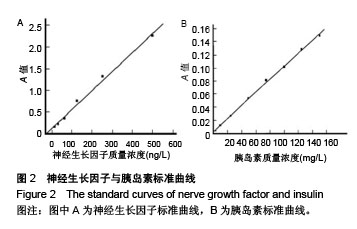
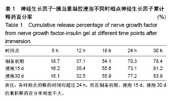
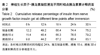
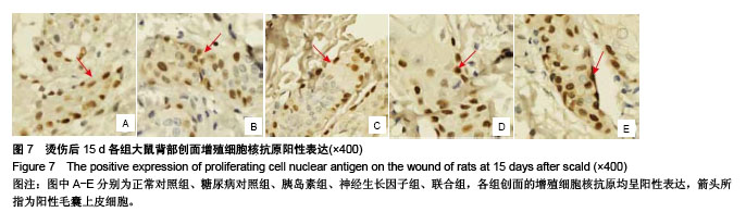
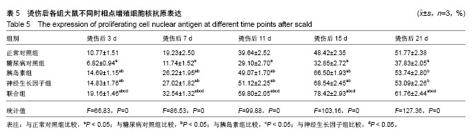
| [1] Chen JC,Lin BB,Hu HW,et al.NGF accelerates cutaneous wound healing by promoting the migration of dermal fibroblasts via the PI3K/Akt-Rac1-JNK and ERK pathways. Biomed Res Int. 2014;2014: 547187.[2] Schenck K,Schreurs O,Hayashi K,et al.The Role of Nerve Growth Factor (NGF) and Its Precursor Forms in Oral Wound Healing.Int J Mol Sci.2017;18(2). pii: E386. doi: 10.3390/ijms18020386.[3] Salvinelli F,Frari V,Rocco ML,et al.Enhanced presence of NGF and mast cells number in nasal cavity after autologous stimulation: relation with sensorineural hearing deficit.Eur Rev Med Pharmacol Sci. 2015; 19(3):381-91.[4] 熊华花,李泉水,范锟.一种新型ngf缓释系统的制备[J].实用临床医学, 2008,9(2):3-6.[5] Okon UA,Umoren IU.Comparison of antioxidant activity of insulin, Ocimum gratissimum L. and Vernonia amygdalina L.in type 1 diabetic rat model.J Integr Med.2017;15(4):302-309. [6] 赵亚男,刘明,张玥,等.糖尿病难愈性创面大鼠模型的建立[J].山东医药, 2016,56(14):31-32.[7] 段强,王冰水,牟翔,等.低功率氦氖激光对小鼠烫伤创面血管内皮细胞因子和细胞凋亡因子基因表达的影响[J].中国康复医学杂志, 2016,31(2): 199-200,214.[8] Pareek G,Shevchuk M,Armenakas NA,et al.The effect of finasteride on the expression of vascular endothelial growth factor and microvessel density: a possible mechanism for decreased prostatic bleeding in treated patients.J Urol. 2003;169(1):20-23.[9] 张雨.烧伤创面修复技术的研究进展[J].医疗装备, 2017,30(10):191-192.[10] 孙晓芳,谢挺.创面修复中生长因子研究及临床干预新进展[J].创伤外科杂志,2016,18(3):190-193.[11] 常清璇,杨沛瑯,刘琰,等.局部应用胰岛素调控基质金属蛋白酶对糖尿病创面血管新生的影响[J].上海交通大学学报(医学版), 2015,35(9):1258.[12] 雷雨.小剂量局部胰岛素联合应用重组人表皮生长因子对烧伤大鼠创面MIP-2和MCP-1表达的影响[J].中国现代医学杂志, 2015,25(6):28-33.[13] 杨沛瑯,常清璇,章雄,等.局部应用胰岛素对糖尿病大鼠烫伤创面愈合的影响[J].中华损伤与修复杂志:电子版,2014,9(4):12-15.[14] 王敏,薛晓东,谢沛霖,等.局部应用NGF-胰岛素复合凝胶对糖尿病大鼠深Ⅱ度烫伤创面修复的影响[J].中国修复重建外科杂志, 2013,27(2):182-188.[15] 杨宗城.烧伤治巧学[M].3版.北京:人民卫生出版化,2006:77-116.[16] Achi NK,Ohaeri OC,Ijeh II,et al.Eleazu C(3).Modulation of the lipid profile and insulin levels of streptozotocin induced diabetic rats by ethanol extract of Cnidoscolus aconitifolius leaves and some fractions: Effect on the oral glucose tolerance of normoglycemic rats.Biomed Pharmacother.2016; 86:562-569.[17] 吴健,薛晓东,刘俊玲,等.胰岛素局部应用对老龄糖尿病大鼠烫伤创面愈合的实验研究[J].中国修复重建外科杂志, 2009,23(12):1482-1486.[18] 李超飞,刘琰,陈雪莲,等.局部应用胰岛素对糖尿病小鼠创面愈合的影响[J].中华损伤与修复杂志:电子版, 2012,7(1):54-61.[19] 肖瑶, 吴基良. 糖尿病引发内皮细胞功能紊乱的研究进展[J].湖北科技学报(医学版),2015, 29(2):177-180.[20] 宋振强,王润秀,于德民,等. 晚期糖基化终末产物修饰人血清白蛋白对离体角质形成细胞迁移功能的影响[J].中华医学杂志, 2008,88(38): 2690-2694.[21] 杨秀颖,张莉,陈熙,等.2型糖尿病周围神经病变机制研究进展[J].中国药理学通报,2016,32(5):598-602.[22] 聂发传,石英.糖尿病周围神经病变发生机制研究进展[J].重庆医学, 2015, 44(1):122-125.[23] 徐江平,郭海彪,汪海涛,等.鼠神经生长因子对糖尿病大鼠皮肤损伤的促愈合作用[J].湖北理工学院学报,2015,31(3):51-56.[24] 李政懋,吕辰,杨选鑫,等.外源NGF促进大鼠皮肤创面模型损伤修复的机制研究[J].上海医药, 2014,35(17):51-53.[25] 曾勇,简华刚,高兵,等.胰岛素外用对糖尿病大鼠深Ⅱ度烧伤创面HIF-1α及VEGF表达的影响[J].重庆医科大学学报, 2010,35(6):860-864.[26] 刘波,王甲汉,韩兆峰.局部胰岛素湿敷对兔深Ⅱ度烫伤创面愈合影响的实验研究[J].南方医科大学学报, 2008,28(4):561-563.[27] 张冀北,薛晓东,谢沛霖,等.局部联合应用NGF及胰岛素对糖尿病大鼠烫伤创面血管形成及Bcl-2、Bax表达的影响[J].中国修复重建外科杂志, 2011,25(3):354-360.[28] 倪向梅.卡波姆980流变学性质及影响因素初探[J].中国科技纵横, 2015, 14(15):201-203.[29] Tseng SY,Chao TH,Li YH,et al.Cilostazol improves high glucose-induced impaired angiogenesis in human endothelial progenitor cells and vascular endothelial cells as well as enhances vasculoangiogenesis in hyperglycemic mice mediated by the adenosine monophosphate-activated protein kina.J Vas Surg. 2016; 63(4):1051-1062.e3.[30] Tseng SY,Chao TH,Li YH,et al.Cilostazol improves high glucose-induced impaired angiogenesis in human endothelial progenitor cells and vascular endothelial cells as well as enhances vas. J Vas Surg.2015;63(4):1051.[31] 王珂,李荣贵,程殿林.增殖细胞核抗原(PCNA)功能及晶体结构研究进展[J].山东轻工业学院学报(自然科学版), 2013,27(3):25-31.[32] 宋楠萌,桑建利,徐恒.增殖细胞核抗原(PCNA)的分子结构及其生物学功能研究进展[J].自然科学进展, 2006,16(10):1201-1209.[33] Lu S.Burn Wound Healing[M]// Chinese Burn Surgery. Springer Netherlands,2015:207-248.[34] Graiani G,Emanueli C,Desortes E,et al.Nerve growth factor promotes reparative angiogenesis and inhibits endothelial apoptosis in cutaneous wounds of Type 1 diabetic mice. Diabetologia. 2004;47(6):1047-1054.[35] Zeng M,Zhi Y,Liu W,et al.Clinical study on local application of low-dose insulin for promoting wound healing after operation for deep burns.Exp Ther Med.2016;12(5):3221-3226.[36] Wang C,Wang J,Feng J.Local application of low-dose insulin in improving wound healing after deep burn surgery.Exp Ther Med. 2016; 12(4):2527-2530.[37] Zhang XJ,Wu X,Wolf SE,et al.Local insulin-zinc injection accelerates skin donor site wound healing.J Surg Res. 2007;142(1):90-96. [38] 谢晓繁,贾赤宇.胰岛素在烧伤创面愈合中的作用[J].中华创伤杂志, 2006, 22(6):475-477.[39] 刘虎仙,贾赤宇.胰岛素及血糖调控与烧伤创面愈合[J].中华烧伤杂志, 2005,21(3):232-234 |
.jpg)


.jpg)