| [1] 张艳苹,沈志强.骨质疏松相关信号通路的研究进展[J].中华骨科杂志, 2017, 37(1):59-64.[2] 陈洁,何跃,兰由玉,等.改良组织块法培养小鼠成骨细胞的研究[J].四川动物,2013, 32(2):283-288.[3] 蒋鹏,宋科官. 破骨细胞及其分化调节机制的研究进展[J]. 中国骨与关节杂志, 2017, 6(3):223-227.[4] 曾展鹏,周驰,李康活,等. 补肾接骨中药对骨折修复的成骨作用及机制[J]. 中国组织工程研究, 2015, 19(15):2442-2448.[5] Huang B, Wang Y, Wang W, et al. mTORC1 Prevents Preosteoblast Differentiation through the Notch Signaling Pathway. Plos Genetics.2015;11(8):e1005426.[6] 范文斌,包倪荣,赵建宁. PI3K/Akt信号通路在骨代谢中的作用[J]. 中国矫形外科杂志, 2013, 21(19):1958-1962.[7] 滕晓英,王连唐.破骨细胞功能调控与骨质疏松症[J]. 中国病理生理杂志, 2002, 18(8):1012-1015.[8] 张苗,张玲莉,雷乐,等. 机械刺激对成骨细胞影响的研究进展[J]. 中国骨质疏松杂志, 2017, 23(9):1240-1244.[9] 朱莎. 骨髓间充质干细胞定向诱导前体骨细胞治疗兔骨损伤的研究[D]. 昆明医科大学, 2012.[10] 张静如,逄丹丹,戴生明. 成骨细胞分化及调节过程[J]. 中华风湿病学杂志, 2016, 20(7):486-488.[11] 刘京利. 未羧化骨钙素促进高糖环境下成骨细胞增殖和分化的机制研究[D]. 中国科学院大学, 2015.[12] 刘中博. Runx2能诱导Osterix在非成骨细胞内的基因表达[D]. 东北师范大学, 2005.[13] 孙强,邱勇. 转录因子Runx2、Osterix与骨髓间质干细胞的成骨分化[J]. 江苏医药, 2006, 32(7):657-659.[14] Dai Q, Xu Z, Ma X, et al. mTOR/Raptor signaling is critical for skeletogenesis in mice through the regulation of Runx2 expression. Cell Death Differ. 2017;24(11):1886-1899.[15] 塔娜,谭明生,移平,等.异补骨脂素对小鼠胚胎前成骨细胞胶原的影响[J].中国中医药信息杂志,2011, 18(4):23-25.[16] 康夏,康菲,杨波,等. miRNA-144靶向调节钙粘蛋白11对间充质干细胞成骨分化的抑制效应[J]. 第三军医大学学报, 2013,35(10): 922-926.[17] Eskildsen T,Taipaleenmäki H,Stenvang J,et al. MicroRNA-138 regulates osteogenic differentiation of human stromal (mesenchymal) stem cells in vivo. Proc Natl Acad Sci U S A. 2011;108(15):6139-6144.[18] Tu XM, Gu YL, Ren GQ. miR-125a-3p targetedly regulates GIT1 expression to inhibit osteoblastic proliferation and differentiation. Exp Ther Med. 2016;12(6):4099-4106.[19] Lee Y,Kim HJ,Park CK,et al.MicroRNA-124 regulates osteoclast differentiation. Bone.2013;56(2):383-389.[20] 王瀚. miR-33-5p在小鼠失重性骨质丢失中的作用及其机制研究[D]. 第四军医大学, 2016.[21] Kureel J, John A A, Dixit M, et al. MicroRNA-467g inhibits new bone regeneration by targeting Ihh/Runx-2 signaling. Int J Biochem Cell Biol. 2017;85:35-43. [22] 唐欢,许海甲,侯煜东,等. Runx2基因对骨代谢调控的研究进展[J]. 中国骨质疏松杂志, 2014,20(12):1501-1505.[23] 李庆庆,黄江鸿,何美健,等. Runx2及其在骨组织工程中的应用[J]. 国际骨科学杂志, 2014, 35(1):44-46.[24] 王钦,李辉,张磊,等. 小鼠颅骨成骨细胞的分离培养及生物学鉴定[J]. 基因组学与应用生物学, 2008, 27(4):325-328.[25] 符毓豪,王菊,周宗瑶. 小鼠成骨细胞体外分离培养及鉴定[J]. 农垦医学, 2008, 30(6):455-458.[26] 陈洁,何跃,兰由玉,等. 改良组织块法培养小鼠成骨细胞的研究[J]. 四川动物, 2013, 32(2):283-288.[27] 郭清皓,蔡钧,杨世疆,等. MicroRNA-203抑制骨肉瘤细胞增殖和迁移的机制研究[J]. 中国现代医学杂志, 2017, 27(6):32-37.[28] 陈斌,符勇,夏雪,等. MicroRNA-638在骨肉瘤中表达下调及对其细胞增殖的影响[J]. 中南医学科学杂志, 2017, 45(4):341-345.[29] 王跃春,张洹,谭广销. 人胎肾间充质样干细胞分离鉴定及其向脂肪和成骨细胞的分化[J]. 中国组织工程研究, 2006, 10(25):17-20.[30] 李静,周国顺,戴利成. 一种骨髓间充质干细胞在体外向成骨细胞分化的诱导方法及诱导培养基:CN 103667182 A[P]. 2014.[31] 宋利格,张秀珍,张克勤,等. 雌激素受体介导的 ERK、JNK 途径在淫羊藿促成骨细胞增殖分化过程中的分子机制[J]. 中华内分泌代谢杂志, 2015, 31(2):148-154.[32] 王潇丽,徐丽丽,杨乃龙.尿酸对人骨髓间充质干细胞成骨分化过程中Wnt信号通路的影响[J].中国组织工程研究,2015,19(28): 4472-4477.[33] 周建,马慧萍,陈克明,等. 50Hz1.8mT电磁场对成骨细胞增殖与分化成熟影响的波形比较研究[J]. 生物化学与生物物理进展, 2011, 38(10):967-974.[34] 闫娟丽,陈克明,周建,等. 脉冲电磁场与正弦交变电磁场对成骨细胞增殖与成熟矿化的比较研究[J]. 解放军医药杂志, 2015,27(3):6-10.[35] 李东伟,刘洪瑜,章孝荣. 哺乳动物早期发育相关microRNAs的研究进展[J]. 生理科学进展, 2013, 44(1):35-38.[36] 张毅,李京玉,廖永晖,等. 血清miRNA:一种新的疾病检测标记物[J]. 中华实验外科杂志, 2011, 28(3):479-480.[37] 张帆,崔庆华. microRNA与人类疾病关系研究中的生物信息学方法和资源[J]. 生理科学进展, 2016,47(3):203-209.[38] 徐兴伟,嵇武. microRNA-155的研究进展[J].中国免疫学杂志, 2012, 28(5):470-473.[39] 冯伟,张幼怡. MicroRNA基因及其靶位点的单核苷酸多态性与疾病易感性[J]. 生理科学进展, 2010, 41(6):443-445.[40] Yang Y,Peng W,Tang T,et al.MicroRNAs as promising biomarkers for tumor-staging:Evaluation of miR21 miR155 miR29a and miR92a in predicting tumor stage of rectal cancer. Asian Pac J Cancer Prev.2014;15(13): 5175-5180.[41] Sun K, Lai E C. Adult-specific functions of animal microRNAs. Nature Reviews Genetics.2013;14(8):535-548.[42] 廖彩秀,桂娅君,肖乾凤,等. MicroRNA-1的心律失常调控作用[J]. 中国动脉硬化杂志, 2015, 23(7):736-739.[43] 张建芳,李建华, 郭军红,等. 卵巢miRNA的表达与功能研究进展[J]. 中国比较医学杂志, 2013, 23(8):67-71.[44] 张学勇,骆学农,郑亚东. microRNA在免疫细胞中作用的研究进展[J]. 细胞与分子免疫学杂志, 2015, 31(5):704-707.[45] 阮正上, 江来. 脓毒症相关的microRNA的研究进展[J]. 医学综述, 2016, 22(1):41-44.[46] Huang K, Fu J, Zhou W, et al. MicroRNA-125b regulates osteogenic differentiation of mesenchymal stem cells by targeting Cbf in vitro. Biochimie.2014;102:47-55.[47] 韩俊,郑苏阳,郭杨,等. miRNA调控破骨细胞分化研究进展[J]. 中国骨质疏松杂志, 2015, 21(11):1393-1396.[48] 连俊翔,杜玮,孟姝. 微小RNAs在破骨细胞分化调控中的研究进展[J]. 华西口腔医学杂志, 2015, 33(5):543-547.[49] 元宇,仝晓阳,邹军. miR-214对骨形成的抑制作用[J]. 中国生物化学与分子生物学报, 2017,33(2):133-137.[50] 宋鹏,荆凯,薛建华,等. miR-195对人骨髓间充质干细胞成骨分化的影响[J].中国组织工程研究, 2016, 20(45):6693-6699.[51] 刘红霞,施琼,安利钦,等. miR-155通过下调BMP9/Smad信号通路抑制间充质干细胞C3H10T1/2成骨分化[J].中国细胞生物学学报, 2017,39(4):410-418.[52] Wang X, Guo B, Li Q, et al. miR-214 targets ATF4 to inhibit bone formation. Nature Medicine. 2013;19(1):93-100.[53] Sugatani T,Vacher J,Hruska KA.A microRNA expression signature of osteoclastogenesis. Blood.2011;117(13):3648.[54] Liu T,Qin AP,Liao B,et al.A novel microRNA regulates osteoclast differentiation via targeting protein inhibitor of activated STAT3 (PIAS3). Bone. 2014; 67(13):156.[55] 邓展涛,金洁雯. miRNA与骨代谢[J].中国矫形外科杂志, 2016, 24(11):1019-1022.[56] Wang Y, Li L, Moore B T, et al. MiR-133a in Human Circulating Monocytes: A Potential Biomarker Associated with Postmenopausal Osteoporosis. Plos One.2012;7(4):e34641. |
.jpg) 文题释义:
成骨前体细胞:成骨细胞根据分化的不同阶段通常可以分为间充质干细胞-未成熟的成骨前体细胞-成熟的成骨细胞-骨细胞。成熟的成骨细胞主要表达Osteocalcin,未成熟的成骨前体细胞主要表达表达Runx-2和Osterix,研究表明未成熟的成骨前体细胞功能非常活跃,其增殖和分化对骨再生非常重要。
Runx-2:是骨髓间充质干细胞成骨分化和骨发育所必需的关键因子,是成骨细胞分化早期最重要的标志基因。
文题释义:
成骨前体细胞:成骨细胞根据分化的不同阶段通常可以分为间充质干细胞-未成熟的成骨前体细胞-成熟的成骨细胞-骨细胞。成熟的成骨细胞主要表达Osteocalcin,未成熟的成骨前体细胞主要表达表达Runx-2和Osterix,研究表明未成熟的成骨前体细胞功能非常活跃,其增殖和分化对骨再生非常重要。
Runx-2:是骨髓间充质干细胞成骨分化和骨发育所必需的关键因子,是成骨细胞分化早期最重要的标志基因。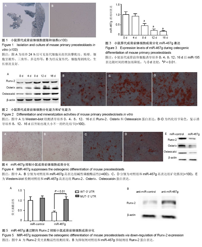