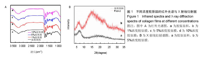
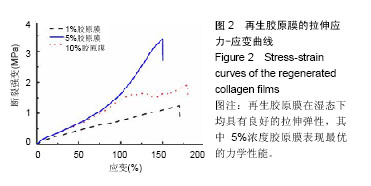
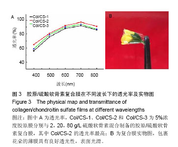
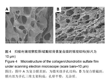
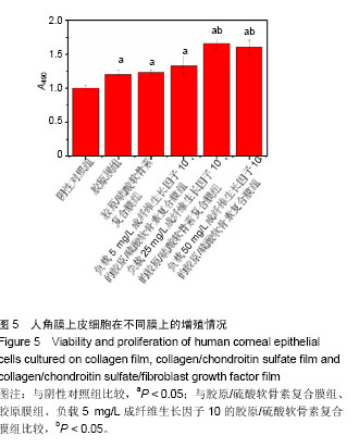
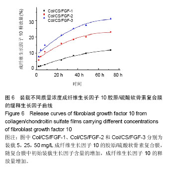
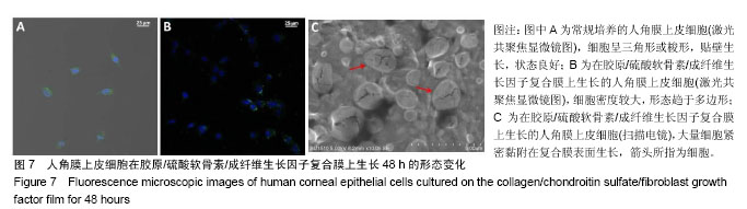
| [1] Mirabelli P,Mukwaya A,Lennikov A,et al.Genome-wide expression differences in anti-Vegf and dexamethasone treatment of inflammatory angiogenesis in the rat cornea.Sci Rep.2017;7(1):7616.[2] Meek KM,Knupp C.Corneal structure and transparency.Prog Retin Eye Res.2015;49(2):1-16.[3] Stafiej P,Kung F,Thieme D,et al.Adhesion and metabolic activity of human corneal cells on PCL based nanofiber matrices. Mater Sci Eng C Mater Biol Appl.2017;71:764-777.[4] Riau AK,Mondal D,Yam GHF,et al.Surface modification of PMMA to improve adhesion to corneal substitutes in a synthetic core-skirt keratoprosthesis.ACS Appl MaterInterfaces.2015;7(39): 21690-21702.[5] Hemalatha T,Yadav S,Krithiga G,et al.Chitosan as a matrix for grafting methyl methacrylate: synthesis, characterization and evaluation of grafts for biomedical applications.Polym Bull(Berl). 2016;73(11): 3105-3117.[6] Yawon H,Gonhyung K.Evaluation of stability and biocompatibility of PHEMA-PMMA keratoprosthesis by penetrating keratoplasty in rabbits. Lab Anim Res.2016;32(4):181-186.[7] 李明.多孔n-HA/PVA/CF(PEEK)复合水凝胶的制备与性能研究[D].深圳:深圳大学化学与化工学院,2015:1-86.[8] Kharaghani D,Meskinfam M,Rezaeikanavi M,et al.Synthesis and characterization of hybrid nanocomposite via biomimetic method as an artificial cornea.Invest Ophth Vis Sci.2015;56(7):5024.[9] Chaiyasan W,Praputbut S,Kompella UB,et al.Penetration of mucoadhesive chitosan-dextran sulfate nanoparticles into the porcine cornea.Colloids Surf B Biointerfaces.2017;149:288-296.[10] Huang X,Wang R,Lu T,et al.Heparin-like chitosan hydrogels with tunable welling behavior, prolonged clotting times, and prevented contact activation and complement activation.Biomacromolecules. 2016;17(12):4011-4020.[11] Jamard M,Hoare T,Sheardown H.Nanogels of methylcellulose hydrophobized with N-tert-butylacrylamide for ocular drug delivery. Drug Deliv Transl Res.2016;6(6):648-659.[12] Tummala GK,Joffre T,Lopes VR,et al.Hyperelastic nanocellulose- reinforced hydrogel of high water content for ophthalmic applications. ACS Biomater Sci Eng.2016;2(11):2072-2079.[13] Sepulveda RV,Valente FL,Reis ECC,et al.Bacterial cellulose and bacterial cellulose/polycaprolactone composite as tissue substitutes in rabbits' cornea.Pesquisa Vet Brasil.2016;36:986-992.[14] Patchan MW,Chae JJ,Lee JD,et al.Evaluation of the biocompatibility of regenerated cellulose hydrogels with high strength and transparency for ocular applications.J Biomater Appl. 2015;30(7):1049-1059.[15] Rudisill SG,DiVito MD,Hubel A,et al.In vitro collagen fibril alignment via incorporation of nanocrystalline cellulose.Acta Biomaterialia.2015; 12(1):122-128.[16] Zhao X,Song W,Liu S,et al.Corneal regeneration by utilizing collagen based materials.Sci China-Chem.2016;59(12):1548-1553.[17] Mohamed-Noriega K,Butron-Valdez K,Vazquez-Galvan J,et al.Corneal melting after collagen cross-linking for keratoconus in a thin cornea of a diabetic patient treated with topical nepafenac: a case report with a literature review.Case Rep Ophthalmol.2017;7(1):119-124.[18] Zhao X,Long K,Liu Y,et al.To prepare the collagen-based artificial cornea with improved mechanical and biological property by ultraviolet-A/riboflavin crosslinking.J Appl Polym Sci.2017;134(38): 45226.[19] Medeiros CS,Giacomin NT,Bueno RL,et al.Accelerated corneal collagen crosslinking: Technique, efficacy, safety, and applications.J Cataract Refract Surg.2016;42(12):1826-1835.[20] Hu C,Yu L,Wei M.Biomimetic intrafibrillar silicification of collagen fibrils through a one-step collagen self-assembly/ silicification approach.RSC Adv.2017;7(55):34624-34632.[21] 刘洁,赵世玉,徐洲,等.胶原在两种离子液体[BMIM]Cl 和[BMIM]Ac 中的溶解及再生结构比较[J].化工学报,2015,66(6):2196-2204.[22] Li C,Liao H,Zhang X,et al.Preparation of cationic modified collagen extracted from leather wastes and their application in dye flocculation.J Appl Polymr Sci.2017;134:45363.[23] Younesi M,Donmez BO,Islam A,et al.Heparinized collagen sutures for sustained delivery of PDGF-BB: delivery profile and effects on tendon-derived cells in-vitro.Acta Biomaterialia.2016;41:100-109.[24] Hariya T,Tanaka Y,Yokokura S,et al.Transparent, resilient human amniotic membrane laminates for corneal transplantation.Biomaterials. 2016;101:76-85.[25] Chae JJ,Choi JS,Lee JD,et al.Physical and biological characterization of the gamma-irradiated human cornea.Cornea. 2015;34(10):1287-1294.[26] Pospichal R,Nesmerak K,Rychlovsky P,et al.Determination of chondroitin sulfate by thiazine dyes using flow injection analysis with spectrophotometric detection.Anal Lett.2007;40(6):1167-1175.[27] Ribeiro AM,Figueiras A,Veiga F.Improvements in topical ocular drug delivery systems: hydrogels and contact lenses.J Pharm Pharm Sci. 2015;18(5):683-695.[28] Cai J,Dou G,Zheng L,et al.Pharmacokinetics of topically applied recombinant human keratinocyte growth factor-2 in alkali-burned and intact rabbit eye.Exp Eye Res.2015;136:93-99. |
.jpg)
.jpg)