1.1 试验设计 前瞻性、单中心、诊断性试验。
1.2 试验完成地点 在中国,广州市正骨医院完成。
1.3 招募 在医院院内公告板张贴研究招募启事招募患者,经过入组标准筛选后,方可纳入研究。研究医生将按照诊疗计划对患者进行治疗及定期的随访。在试验期间,医院将提供一定的随访车费补贴,而且随访阶段免除挂号费和实验室检查费用。
1.4 受试者选择 选取广州市正骨医院门诊就诊及住院治疗的踝关节扭伤患者共70例。
(1)试验组纳入标准:
•踝关节扭伤后1-7 d患者;
•需进行踝关节副韧带损伤修复手术者;
•年龄16-42岁者。
(2)试验组排除标准:
•行MRI和超声检查间隔超过3 d以上的患者;
•合并踝关节感染者;
•合并踝骨骨折者。
(3)对照组纳入标准:为患者未损伤侧作磁共振检查。
1.5 试验分组及盲法 采用患者健侧和患侧影像检查数据对比的方法,进行自身对照比较,试验未采用随机化和盲法分组。
1.6 高频超声检查
1.6.1 检查仪器 采用柯尼卡美能达医疗器材印刷有限公司彩超机7-14 MHz探头进行超声监测,采用美国GE公司的GE signa HDX 3.0T磁共振仪进行MRI检测。以MRI检测或手术结果判断的韧带损伤为诊断金标准。
1.6.2 检查方法
(1)检查外踝韧带:首先检查距腓前韧带,如果患者体位允许,将患肢稍内旋放于检查床上,充分暴露外踝,探头首先放置在腓骨远段外侧,向下移动直至腓骨尖部。探头远端向上,向前移动以显示距骨。此处距腓前韧带由于斜向走形而常出现各向异性伪像,超声上显示为回声均匀的低回声结构。如果将探头一端轻抬,另一端轻压,使韧带纤维垂直于声束以消除各向异性伪像,则正常距腓前韧带长轴切面可显示为连续的、内部呈致密纤维状的带状高回声结构,自腓骨延伸至距骨。
(2)检查距腓前韧带:利用韧带的各向异性伪像对其进行定位,因相对于周围较高回声的脂肪组织,韧带由于各向异性伪像而呈低回声会更容易被显示。当明确为韧带结构后,要尽可能消除此伪像,所以在扫查过程中探头需要沿着腓骨头做旋转,多角度多切面扫查,以正确显示距腓前韧带的声像图特征。
(3)检查跟腓韧带:将探头置于外踝斜冠状切面,其上缘与外踝尖部平齐,其下略微向后,长轴切面表现为一条索状的丝状结构,覆盖跟骨的外侧面,短轴断面显示跟腓韧带为卵圆形,偶尔,可能类似于关节内和腓骨肌腱相关的游离体,跟腓韧带头侧部分走行在腓骨肌腱的深面,因各向异性而呈低回声。
检查时一定需做扇形扫查,以充分显示韧带各个层面,跟腓韧带的超声表现与距腓前韧带类似,但是不全一样,它的凹状走形使超声检查其外踝附着点更为困难。注意在探头与外踝后方的凹陷处堆积足量的耦合剂,起到良好的透声垫的作用,便于观察,研究表明改变足的体位可以显示跟腓韧带的全长。
嘱患者做背屈运动,使跟腓韧带被动拉伸,在运动中观察韧带全程,从而更准确的诊断跟腓韧带的损伤情况,特别是在韧带损伤严重时,周围组织模糊不便于观察时这种方法尤为重要。当跟腓韧带发生急性损伤时,同时需要观察其周边的情况,特别是腓骨长短肌腱,以防误诊。在检查跟腓韧带时,如果在踝关节背曲过程中能显示跟腓韧带的带状回声且腓肌腱向外侧移动,就能排除跟腓韧带的完全断裂。
1.6.3 超声和MRI诊断侧副韧带损伤的分级标准
MRl诊断分级[15]:Ⅰ级损伤,韧带完整,仅见围绕韧带的软组织水肿;Ⅱ级损伤,韧带部分撕裂,显示为韧带形态不规则、纤维束部分完整,韧带内显示液体信号;Ⅲ级损伤,韧带完全断裂,即韧带形态消失或连续性中断。
超声诊断分级[16]:挫伤,表现为韧带增厚,回声减低,连续性好;部分撕裂,表现为韧带纹理连续性部分中断或变薄,周边关节腔可有积液;完全断裂,表现为韧带的连续性完全中断,断端回缩,周围关节腔可伴有积液。
1.6.4 术中观察韧带损伤的诊断标准 患者均进行损伤侧外侧副韧带修补手术。根据美国医学会(AMA)标准解剖分类法(https://www.ama-assn.org/),术后探查判断踝关节韧带损伤的标准:Ⅰ度,仅见距腓前韧带部分或完全断裂,即超声的部分撕裂、完全撕裂损伤;Ⅱ度,距腓前韧带和跟腓韧带同时部分或完全撕裂;Ⅲ度,距腓前韧带、跟腓韧带和胫腓前韧带同时部分或完全撕裂。
1.7 结局指标
1.7.1 主要结局指标 入院后1 d高频超声检测踝关节外侧副韧带完全断裂的敏感度:为真阳性率,即实际为踝关节外侧副韧带完全断裂又被诊断标准正确诊断的患者例数百分比。敏感度越高说明漏诊率越低。
1.7.2 次要结局指标
(1)入院后1 d高频超声检测踝关节外侧副韧带挫伤及部分断裂的敏感度:检测方法及标准同上。
(2)入院后1 d高频超声检测踝关节外侧副韧带挫伤、部分断裂、完全断裂的特异度:为真阴性率,就是实际无韧带损伤(健侧)按该诊断标准被正确地判定无韧带损伤的患者例数百分比。特异度越高说明误诊率越低。
(3)入院后1 d高频超声检测踝关节外侧副韧带挫伤、部分断裂、完全断裂阳性预测值:阳性预测值是指高频超声检出的全部阳性(韧带损伤)例数中,真正“韧带损伤”的例数(真阳性)所占的比例,可反映高频超声阳性结果者出现韧带损伤的可能性。
(4)入院后1 d高频超声检测踝关节外侧副韧带挫伤、部分断裂、完全断裂阴性预测值:阴性预测值指高频超声检测结果为阴性(韧带未损伤)的患者中真正韧带未损伤的比例。可反映高频超声阴性结果者未出现韧带损伤的可能性。
(5)入院后1 d高频超声检测踝关节外侧副韧带挫伤、部分断裂、完全断裂诊断正确率:为高频超声检测的真阳性例数和真阴性例数,与总患者数的百分比。数值越高说明诊断越正确。
(6)入院后1 d高频超声检测踝关节外侧副韧带挫伤、部分断裂、完全断裂ROC曲线下面积值:以敏感性为纵坐标代表真阳性率,(1-特异性)为横坐标代表假阳性率,作图绘成ROC曲线。ROC曲线下的面积值为0.5-1.0。在AUC>0.5的情况下,AUC越接近于1,说明诊断效果越好。AUC=0.5时,说明诊断方法无诊断价值。
(7)入院后1 d高频超声检测踝关节外侧副韧带挫伤、部分断裂、完全断裂ROC曲线截断点值:选择最佳的诊断界限值。ROC曲线越靠近左上角,试验的诊断准确性就越高。最靠近左上角的ROC曲线的点是错误最少的最好阈值,其假阳性和假阴性的总数最少。
(8)入院后1 d高频超声检测踝关节外侧副韧带形态的超声成像形态:采用高频超声检测外侧副韧带形态,以判断外侧副韧带损伤的3种类型挫伤、部分断裂、完全断裂的超声形态。
1.8 不良事件 研究者对患者随访时出现的不良反应(具体见次要结果指标所述)做详细记录。随访过程中要详细记录不良反应发生的时间、类型及当时的处理方法,并在24 h内详细报知研究负责人和伦理审查委员会。
1.9 试验流程 试验将纳入来自广州市正骨医院踝关节扭伤并接受踝关节韧带修复手术治疗患者70例,分别进行损伤侧和健侧踝关节高频超声检测,设为试验组和对照组,见图1。
1.10 样本量估算 以往研究结果显示,超声诊断踝关节外侧副韧带损伤的敏感性、特异性分别为84%及93%[17],结合课题组临床经验,预计此次试验高频超声检测踝关节外侧副韧带完全断裂的敏感度和特异度分别为85%和95%,敏感度和特异度容许误差均为10%,设1-α=95%,显著性水准单侧α=0.05,采用PASS 11.0软件(PASS,Kaysville,UT,USA)计算后得知,估算灵敏度(试验组)所需样本量为58例,估算特异度(对照组)所需样本量为29例,遵循每组样本量相等的原则,取上述样本量最大值58例,加上20%的脱落病例,最终总样本量设为70例。
1.11 统计学考虑
(1)数据描述:统计学分析由统计分析人员应用SPSS 20.0统计软件包(IBM,Armonk,NY,USA)完成,且符合意向分析原则。计量资料符合正态分布用均值(mean)、标准差(SD)表示,非正态分布数据用下四分位数(q1)、中位数(median)和上四分位数(q3)表示。计数资料和等级资料用百分率表示。
(2)分析方法:试验组和对照组高频超声检测踝关节外侧副韧带挫伤、部分断裂、完全断裂的敏感度,特异度、阳性预测值、阴性预测值、诊断正确率的比较采用Friedman秩和检验法,试验组和对照组ROC曲线下面积值及截断点值的比较采用配对t 检验(正态分布数据)和(非态分布数据)Wilcoxon配对检验。采用SPSS 20.0软件绘制诊断ROC曲线,计算截断点。检验水准双侧α=0.05。
(3)试验分析的数据集:试验纳入分析的研究人群主要纳入符合方案集:指所完成试验,在全分析集的基础上,符合入选和排除标准、具备有效的基线值、依从性好、并且没有违背临床试验方案,完成病例报告表规定填写内容的受试人群。
1.12 数据收集和管理
(1)数据收集方法:以病例报告表的形式收集数据,并经统一表格汇总。数据以双重录入形式转化为电子版文件。
(2)数据管理:试验将数据库交给专业统计人员进行统计分析,并由统计分析人员写出统计分析报告,交付试验的主要研究者,写出研究报告。试验锁定后的数据文件不允许再作变动并将数据库保存备查。所有与本次临床试验有关的研究资料均由广州市正骨医院保存。
1.13 监控方法
(1)数据监控委员会的组成:数据监控委员会成员具备的资格由所监察的研究的性质所决定。数据监控委员会具备完善的组织,在研究开始前开会审查数据监控委员会章程与研究方案。
(2)研究者资质:此次试验的影像科医生均具有丰富的超声及MRI检查经验,由该专业学科带头人或主任医师、高年资副主任医师承担。试验数据处理和统计计算由统计学专业人员完成。
(3)审核:监查员确认所有数据的记录与报告是否正确完整,所有病例报告表填写是否正确,并与原始资料一致。每一受试者的治疗变更、间发疾病、失访、检查遗漏等均应确认并记录。
(4)补偿:患者明确被医院诊断为患了某种在专项医疗保险合同上列明的疾病或手术,将由保险公司根据合同约定的金额来给付给患者检查、治疗及护理费用。
1.14 伦理与传播 试验经广州市正骨医院医学伦理委员会批准(审批单位:广州市正骨医院医院,审批时间:2018年5月,审批号:2018-05)。研究符合世界医学会制定的《赫尔辛基宣言》的要求。参加试验及试验中的个人资料均属保密,将依照法律规定得到保护。研究结果将在同行评议期刊或以会议报告形式发表,出版数据将公开发布于www.figshare.com。
中国组织工程研究杂志出版内容重点:人工关节;骨植入物;脊柱;骨折;内固定;数字化骨科;组织工程
.jpg)
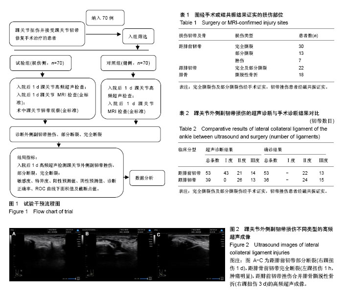
.jpg)