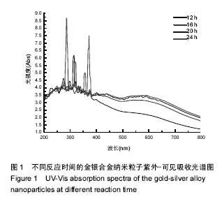
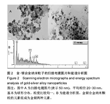
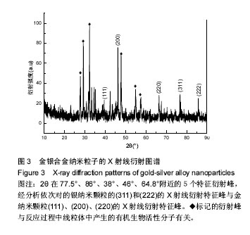
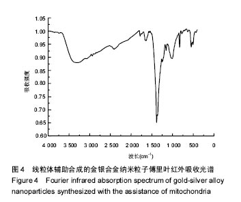
| [1]Huang J,Wang W,Lin L,et al.A general strategy for the biosynthesis of gold nanoparticles by traditional Chinese medicines and their potential application as catalysts.Chem Asian J. 2009;4(7):1050-1054.[2]Ray PC,Darbha GK,Ray A,et al.A gold-nanoparticle-based fluorescence resonance energy transfer probe for multiplexed hybridization detection: accurate identification of bio-agents DNA. Nanotechnology.2007;18:1-6.[3]Schwartzberg AM,Grant CD,Wolcott A,et al.Unique gold nanoparticle aggregates as a highly active surface-enhanced Raman scattering substrate.J Phys Chem B. 2004;108(50): 19191-19197.[4]Pissuwan D,Niidome T,Cortie MB.The forthcoming applications of gold nanoparticles in drug and gene delivery systems.J Controlled Release.2011;149(1):65.[5]Boisselier E,Astruc D.Gold nanoparticles in nanomedicine: preparations, imaging, diagnostics, therapies and toxicity. Chem Soc Rev.2009;38(6):1759-1782. [6]Li Z,Huang P,Zhang X,et al.RGD-Conjugated Dendrimer-Modified Gold Nanorods for in Vivo Tumor Targeting and Photothermal Therapy.Mol Pharm.2009;7(1):94-104. [7]Chen DH,Chen CJ.Formation and characterization of Au–Ag bimetallic nanoparticles in water-in-oil microemulsions.J Mater Chem.2002;12(5):1557-1562.[8]Papavassiliou GC.Surfaceplasmons in small Au-Ag alloy particles.J Phys Metal Phys.1976;6(4):L103.[9]Link S,Wang ZL,El-Sayed MA.Alloy formation of gold-silver nanoparticles and the dependence of the plasmon absorption on their composition.J Phys Chem B.1999;103(18): 3529-3533.[10]Shankar SS,Ahmad A,Pasricha R,et al.Bioreduction of chloroaurate ions by geranium leaves and its endophytic fungus yields gold nanoparticles of different shapes.J Mater Chem.2003;13(7):1822-1826.[11]Shankar S,Ahmad A,Sastry M.Geranium leaf assisted biosynthesis of silver nanoparticles. Biotechnol Prog. 2003; 19(6):1627-1631.[12]Narayanan KB,Sakthivel N.Phytosynthesis of gold nanoparticles using leaf extract of Coleus amboinicusLour.Mater Charact.2010;61:1232-1238.[13]Faramarzi MA,Forootanfar H.Biosynthesis and characterization of gold nanoparticles produced by laccase from Paraconiothyriumvariabile.Colloids Surf B Biointerfaces. 2011;87(1):23-27.[14]Philip D.Honey mediated green synthesis of gold nanoparticles.Spectrochim Acta A Mol Biomol Spectrosc. 2009;73(4):650-653. [15]Nune SK,Chanda N,Shukla R,et al.Green nanotechnology from tea: phytochemicals in tea as building blocks for production of biocompatible gold nanoparticles.J Mater Chem.2009;19(19):2912-2920.[16]Katz E,Willner I,Wang J.Electroanalytical and bioelectroanalytical systems based on metal and semiconductor nanoparticles.Electroanalysis. 2004;16(1-2): 19-44.[17]Tisch U,Haick H.Arrays of chemisensitive monolayer-capped metallic nanoparticles for diagnostic breath testing.Rev Chem Eng.2011;26(5-6):171-179.[18]Ahn H,Chandekar A,Kang B,et al.Electrical conductivity and vapor-sensing properties of ω-(3-thienyl) alkanethiol-protected gold nanoparticle films.Chem Mater. 2004;16(17):3274-3278.[19]Wang AQ,Chang CM,Mou CY.Evolution of catalytic activity of Au-Ag bimetallic nanoparticles on mesoporous support for CO oxidation.J Phys Chem B.2005;109(40):18860-18867.[20]Mallin MP,Murphy CJ.Solution-phase synthesis of sub-10 nm Au-Ag alloy nanoparticles.Nano Lett.2002;2(11):1235-1237.[21]Endo T,Yoshimura T,Esumi K.Synthesis and catalytic activity of gold silver binary nanoparticles stabilized by PAMAM dendrimer.J Colloid Interface Sci.2005;286(2):602-609.[22]Boisselier E,Astruc D.Gold nanoparticles in nanomedicine: preparations, imaging, diagnostics,therapies and toxicity. Chem Soc Rev.2009;38(6):1759-1782.[23]Bond GC. Gold: a relatively new catalyst. Catal Today. 2002;72(1-2): 5-9.[24]Li Z,Huang P,Zhang X,et al.RGD-conjugated dendrimer- modified gold nanorods for in vivo tumor targeting and photothermal therapy.Mol Pharm.2010;7(1):94-104.[25]Cu D,Zhang H,Wang K,et al.Gold Nanoparticles Enhance Efficiency of In Vitro Gene Transcription-Translation System.Nano Biomed Eng.2011;3(2):120-125.[26]Gole A, Murphy CJ.Seed-mediated synthesis of gold nanorods: role of the size and nature of the seed.Chem Mater.2004;16(19):3633-3640. [27]Meltzer S,Resch R,Koel BE,et al.Fabrication of nanostructures by hydroxylamine seeding of gold nanoparticle templates.Langmuir.2001;17(5):1713-1718. [28]Westcott SL,Oldenburg SJ,Lee TR, et al.Formation and adsorption of clusters of gold nanoparticles onto functionalized silica nanoparticle surfaces. Langmuir. 1998;14(19):5396-5401. [29]Ahmad A,Senapati S,Khan MI,et al.Extracellular biosynthesis of monodisperse gold nanoparticles by a novel extremophilic actinomycete, Thermomonospora sp. Langmuir.2003;19(8):3550-3553. [30]Mukherjee P,Senapati S,Mandal D,et al.Extracellular synthesis of gold nanoparticles b the fungus Fusarium oxysporum.Chembiochem.2002;3(5):461-463. [31]Xie J,Lee JY,Wang DIC,et al.High-yield synthesis of complex gold nanostructures in a fungal system. J Phys Chem C. 2007;111(45):16858-16865. |
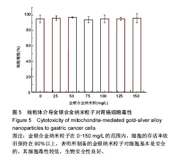
.jpg)
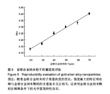
.jpg)