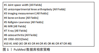
[1] HADA S, ISHIJIMA M, KANEKO H, et al. Association of medial meniscal extrusion with medial tibial osteophyte distance detected by T2 mapping MRI in patients with early-stage knee osteoarthritis. ArthritiRes Ther. 2017;19(1):201-212.
[2] SEKIYA, I, SASAKI, S, MIURA, Y, et al. Medial ibial osteophyte width strongly reflects medial meniscus extrusion distance and medial joint space width moderately reflects cartilage thickness in knee radiographs.J Magn Reson Imaging. 2022;56(3):824-834.
[3] BLOECKER K, WIRTH W, HUNTER DJ, et al. Contribution of regional 3D meniscus and cartilage morphometry by MRI to joint space width in fixed flexion knee radiography-a between-knee comparison in subjects with unilateral joint space narrowing. Eur J Radiol. 2013; 82(12):e832-e839.
[4] HUNTER DJ, ZHANG YQ, TU X, et al. Change in joint space width: hyaline articular cartilage loss or alteration in meniscus. Arthritis Rheum. 2006; 54(8):2488-2495.
[5] PARSONS CM, JUDGE A, MEYER R, et al.Determining individual trajectories of joint space loss: improved statistical methods for monitoring knee osteoarthritis disease progression. Osteoarthritis Cartilage. 2021;29(1): 59-67.
[6] HUNTER DJ, BUCK R, VIGNON E, et al. Relation of regional articular cartilage morphometry and meniscal position by MRI to joint space width in knee radiographs. Osteoarthritis Cartilage. 2009;17(9):1170-1176.
[7] ADAMS JG, MCALINDON T, DIMASI M, et al. ontribution of meniscal extrusion and cartilage loss to joint space narrowing in osteoarthritis. Clin Radiol. 1999;54(8):502-506.
[8] BEATTIE KA, DURYEA J, PUI M, et al. Minimum joint space width and tibial cartilage morphology in the knees of healthy individuals: a cross-sectional study. BMC Musculoskelet Disord. 2008;9:119.
[9] ROTH M, EMMANUEL K, WIRTH W, et al.Sensitivity to change and
association of three-dimensional meniscal measures with radiographic joint space width loss in rapidclinical progression of knee osteoarthritis.Eur Radial. 2018;28(5):1844-1853.
[10] ELLIS RT, NETTROUR JF, KEENEY JA. TKA is More Durable Than UKA for Morbidly Obese Patients: A Two-Year Minimum Follow-Up Study. Arthroplasty. 2021;36(6):1933-1941.
[11] O’NEILL CN, GOWD AK, WATERMAN BR, et al. Significant Reduction in Short-Term Complications Following Unicompartmental Versus Total Knee Arthroplasty: A Propensity Score Matched Analysis. Arthroplasty. 2022;37(10):2014-2019.
[12] KLASAN A, TAY ML, FRAMPTON C, et al. High usage of medial unicompartmental knee arthroplasty negatively influences total knee arthroplasty revision rate.Knee Surg Sports Traumatol Arthrosc. 2022; 30(9):3199-3207.
[13] PONGCHAROEN B, LIENGWATTANAKOL P, BOONTANAPIBUL K. Comparison of Functional Recovery Between Unicompartmental and Total Knee Arthroplasty: A Randomized Controlled Trial. J Bone Joint Surg. 2023;105(3):191-201.
[14] DI MARTINO A, BORDINI B, BARILE F, et al. Unicompartmental knee arthroplasty has higher revisions than total knee arthroplasty at long term follow-up: a registry study on 6453 prostheses. Knee Surg Sports Traumatol Arthrosc. 2021;29(10):3323-3329.
[15] LEE CS, SU EP, CROSS MB, et al. Unicompartmental Knee Arthroplasty Is Associated With a Lower Rate of Periprosthetic Joint Infection Compared to Total Knee Arthroplasty. Arthroplast Today. 2021;10:117-122.
[16] FISHER N, AGARWAL M, REUBEN SF, et al. Sporting and physical activity following Oxford medial unicompartmental knee arthroplasty. Knee. 2006;13(4):296-300.
[17] GOODFELLOW J, O’CONNOR J, DODD C, et al. Unicompartmental arthroplasty with the Oxford knee. New York: Oxford University Press, 2006: 44-45,169.
[18] NIINIMÄKI TT, MURRAY DW, PARTANEN J, et al.Unicompartmental knee arthroplasties implanted for osteoarthritis with partial loss of joint space have high re-operation rates. Knee. 2011;18(6):432-435.
[19] PANDIT H, GULATI A, JENKINS C, et al. Unicompartmental knee replacement for patientswith partial thickness cartilage loss in the affected compartment. Knee. 2011;18(3):168-171.
[20] MAIER MW, KUHS F, STREIT MR, et al. Unicompartmental knee arthroplasty in patients with full versus partial thickness cartilage loss (PTCL): equal in clinical outcome butwithhigher reoperation rate for patients with PTCL. Arch Orthop Trauma Surg. 2015;135(8):1169-1175.
[21] KNIFSUND J, HATAKKA J, KEEMU H, et al. Unicompartmental knee arthroplasties are performed on the patients with radiologically too mild osteoarthritis.ScandJ Surg. 2017;106(4):338-341.
[22] HAMILTON TW, PANDIT HG, INABATHULA A, et al. Unsatisfactory outcomes following unicompartmental knee arthroplasty in patients with partial thickness cartilage loss: a medium-term follow-up. Bone Joint J. 2017;99-B(4):475-482.
[23] WURM A, ZECHLING A, LEITNER H, et al. Medial unicondylar knee arthroplasty should be reserved for patients with complete joint space collapse. Knee Surg Sports Traumatol Arthrosc. 2022; 30(9):3162-3167.
[24] CARLSON SW, VARGAS-HERNANDEZ JS, CARLSON BC, et al. Lack of bone-on-bone arthritis is not a strict contraindication for mobile-bearing unicompartmental knee arthroplasty. JBJS Open Access. 2020; 5(3):e19.00058-e19.00058.
[25] WONG KC, LEE M, LIOW L, et al. Bone-on-bone contact on radiograph is not a prerequisite for successful outcome in fixed-bearing medial unicompartmental kneearthroplasty-A 10-year follow-up study. J Knee Surg. 2023;36(6):658-666.
[26] NIINIMAKI T, ESKELINEN A, MAKELA K, et al. Unicompartmental knee arthroplasty survivorship is lower than TKA survivor-ship:A 27-year finnish registry study. Clin Orthop Relat Res. 2014;472(5):1496-1501.
[27] 李世超,解光越,孙振,等.应力位X射线片与单髁置换后膝关节对位对线的关系[J].中国组织工程研究,2023,27(18):2910-2914.
[28] VASSO M, DEL REGNO C, D’AMELIO A, et al.Minor varus alignment provides better results than neutral alignment in medial UKA. Knee. 2015;22(2):117-121.
[29] YUE J, GUO W, WAN F, et al. Quantitative Evaluation of joint space width in the lateral compartment after medial unicompartmental knee arthroplasty: comparison of three radiographic views. J Knee Surg. 2018; 31(8):730-735.
[30] KHAMAISY S, ZUIDERBAAN HA, VAN DER LIST JP, et al. Medial unicompartmental knee arthroplasty improves congruence and restores joint space width of the lateral compartment.Knee. 2016;23(3):501-505.
[31] 俞颖豪,赵继军,刘冬铖,等. 数字图像术前规划辅助单髁置换对固定平台假体摆位的临床指导意义[J]. 中国组织工程研究,2021,25(21): 3324-3331.
[32] KELLGREN JH, LAWRENCE JS. Radiological assessment of osteoarthrosis. Ann Rheum Dis. 1957;16(4):494-502.
[33] FDA/CDER resource page. Food and Drug Administration Web site. Guidance for industry: clinical development programs for drugs, devices, and biological products intended for the treatment of osteoarthritis. Available at: http://www.fda.gov/cder/guidance/2199dft.htm. Accessed November 28, 2002.
[34] The European Agency for the Evaluation of Medicinal Products Human Medicines Committee for proprietary medicinal products (CTMP) points to consider on the clinical investigation of medicinal products used in the treatment of osteoarthritis. Available at:http://www.eudra.org. Accessed November 28, 2002.
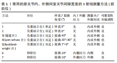
[35] BUCKLAND-WRIGHT JC, MACFARLANE DG, WILLIAMS SA, et al. Accuracy and precision of joint space width measurements in standard and macroradiographs of osteoarthritic knees. Ann Rheum Dis. 1995;54(11): 872-880.
[36] JANSEN MP, MASTBERGEN SC, ECKSTEIN F, et al. Comparison between 2D radiographic weight-bearing joint space width and 3D MRI non-weight-bearing cartilage thickness measures in the knee using non-weight-bearing 2D and 3D CT as an intermediary. Ther Adv Chronic Dis. 2021;12:20406223211037868.
[37] PIPERNO M, HELLIO LE GRAVERAND MP, CONROZIER T, et al. Quantitative evaluation of joint space width in femorotibial osteoarthritis: comparison of three radiographic views. Osteoarthritis Cartilage. 1998;6(4):252-259.
[38] VIGNON E, PIPERNO M, LE GRAVERAND MP, et al. Measurement of radiographic joint space width in the tibiofemoral compartment of the osteoarthritic knee: comparison of standing anteroposterior and Lyon schuss views. Arthritis Rheum. 2003;48(2):378-384.
[39] CHEUNG JC, TAM AY, CHAN LC, et al. Superiority of multiple-joint space width over minimum-joint space width approach in the machine learning for radiographic severity and knee osteoarthritis progression. Biology (Basel). 2021;10(11):1107.
[40] PAIXAO T, DIFRANCO MD, LJUHAR R, et al. A novel quantitative metric for joint space width: data from the Osteoarthritis Initiative (OAI). Osteoarthritis Cartilage. 2020;28(8):1055-1061.
[41] YANAGISAWA S, OHSAWA T, SAITO K, et al. Morphological evaluation and diagnosis of medial type osteoarthritis of the knee using ultrasound. J Orthop Sci. 2014;19(2):270-274.
[42] ZHU J, LI B, QIU L, et al. A measurement method of knee joint space width by ultrasound: a large multicenter study. Quant Imag Med Sug. 2020;10(5):979-987.
[43] ECKSTEIN F, GUERMAZI A, GOLD G, et al. Imaging of cartilage and bone: promises and pitfalls in clinical trials of osteoarthritis. Osteoarthritis Cartilage. 2014;22(10):1516-1532.
[44] PELLETIER JP, COOPER C, PETERFY C, et al. What is the predictive value of MRI for the occurrence of knee replacement surgery in knee osteoarthritis? Ann Rheum Dis. 2013;72(10):1594-1604.
[45] CICUTTINI FM, JONES G, FORBES A, et al. Rate of cartilage loss at two years predicts subsequent total knee arthroplasty: a prospective study. Ann Rheum Dis. 2004;63(9):1124-1127.
[46] RAYNAULD JP, MARTEL-PELLETIER J, HARAOUI B, et al. Risk factors predictive of joint replacement in a 2-year multicentre clinical trial in knee osteoarthritis using MRI: results from over 6 years of observation.Ann Rheum Dis. 2011;70(8):1382-1388.
[47] ECKSTEIN F, BOUDREAU R, WANG Z, et al. Comparison of radiographic joint space width and magnetic resonance-imaging for prediction of knee replacement-A longitudinal case-control study from the osteoarthritis initiative. Eur Radiol. 2016;26(6): 1942-1951.
[48] LE GRAVERAND MP, BUCK RJ, WYMAN BT, et al. Change in regional cartilage morphology and joint space width in osteoarthritis participants versus healthy controls: a multicentre study using 3.0 Tesla MRI and Lyon-Schuss radiography. Ann Rheum Dis. 2010;69(1):155-162.
[49] KOTHARI MD, RABE KG, ANDERSON DD, et al. The relationship of three-dimensional joint space width on weight bearing CT with pain and physical function. J Orthopaed Res. 2019:10.1002/jor.24566.
[50] TURMEZEI TD, BLOW S, RUPRET S, et al. Quantitative three-dimensional assessment of knee joint space width from weight-bearing CT. Radiology. 2021;299(3):649-659.
[51] TURMEZEI TD, LOW SB, RUPRET S, et al. Multiparametric 3-D analysis of bone and joint space width at the knee from weight bearing computed tomography. Osteoarthr Imaging. 2022;2(2):100069. |