[1] SI L, WINZENBERG TM, JIANG Q, et al. Projection of osteoporosis-related fractures and costs in China: 2010-2050. Osteoporos Int. 2015;26(7):1929-1937.
[2] LODOSO-TORRECILLA I, VAN DEN BEUCKEN JJJP, JANSEN JA. Calcium phosphate cements: optimization toward biodegradability. Acta Biomater. 2021;119:1-12.
[3] NASROLLAHI N, DEHKORDI AN, JAMSHIDIZAD A, et al. Preparation of brushite cements with improved properties by adding graphene oxide. Int J Nanomedicine. 2019;14:3785-3797.
[4] SHEIKH Z, ZHANG YL, GROVER L, et al. In vitro degradation and in vivo resorption of dicalcium phosphate cement based grafts. Acta Biomater. 2015;26:338-346.
[5] 彭磊,丁秀明,陈克伟,等.羟基磷灰石体系透钙磷灰石骨水泥的理化性能[J].中国组织工程研究,2018,22(6):821-826.
[6] DING L, WANG H, LI J, et al. Preparation and characterizations of an injectable and biodegradable high-strength iron-bearing brushite cement for bone repair and vertebral augmentation applications. Biomater Sci. 2023;11:96-107.
[7] LU TL, WANG JC, YUAN XY, et al. Zinc-doped calcium silicate additive accelerates early angiogenesis and bone regeneration of calcium phosphate cement by double bioactive ions stimulation and immunoregulation. Biomater Adv. 2022;141:213120.
[8] BOSE S, FIELDING G, TARAFDER S, et al. Understanding of dopant-induced osteogenesis and angiogenesis in calcium phosphate ceramics. Trends Biotechnol. 2013;31(10):594-605.
[9] MIRTCHI AA, LEMAITRE J, TERAO N. Calcium phosphate cements: study of the beta-tricalcium phosphate--monocalcium phosphate system. Biomaterials. 1989;10(7):475-480.
[10] BOHNER M. Calcium orthophosphates in medicine: from ceramics to calcium phosphate cements.Injury. 2000;31 Suppl 4:37-47.
[11] HURLE K, OLIVEIRA JM, REIS RL, et al. Ion-doped brushite cements for bone regeneration. Acta Biomater. 2021;123:51-71.
[12] KANNAN S, GOETZ-NEUNHOEFFER F, NEUBAUER J, et al. Rietveld structure and in vitro analysis on the influence of magnesium in biphasic (hydroxyapatite and beta-tricalcium phosphate) mixtures. J Biomed Mater Res B Appl Biomater. 2009;90B(1):404-411.
[13] LAURA T, VAQUERO M. Chronic iron deficiency as an emerging risk factor for osteoporosis: a hypothesis. Nutrients. 2015;7(4):2324-2344.
[14] BALOGH E, PARAGH G, JENEY V. Influence of iron on bone homeostasis. Pharmaceuticals (Basel). 2018;11(4):107.
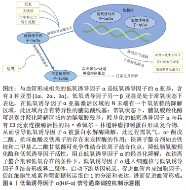
[15] 贾鹏,邓廉夫.低氧诱导因子-α信号通路与骨形成[J].中华骨科杂志,2015,35(6):676-680.
[16] LI G, NAN Z, ZHAO S, et al. Fe-doped brushite bone cements with antibacterial property. Mater Lett. 2017;215:27-30.
[17] VAHABZADEH S, FLECK S, MARBLE J, et al. Role of iron on physical and mechanical properties of brushite cements, and interaction with human dental pulp stem cells. Ceram Int. 2020;46(8 Pt B):11905-11912.
[18] USKOKOVIĆ V, GRAZIANI V, WU V, et al. Gold is for the mistress, silver for the maid: enhanced mechanical properties, osteoinduction and antibacterial activity due to iron doping of tricalcium phosphate bone cements. Mater Sci Eng C Mater Biol Appl. 2019;94:798-810.
[19] SAGHIRI MA, ASATOURIAN A, ORANGI J, et al. Functional role of inorganic trace elements in angiogenesis-Part II: Cr, Si, Zn, Cu, and S. Crit Rev Oncol Hematol. 2015;96(1):143-155.
[20] 郭苏童,郭宇,王凌,等.铜离子在骨组织工程中的应用:生物相容性,抗菌性,血管生成活性及成骨活性[J].中国组织工程研究,2022,26(21):3410-3414.
[21] RYAN EJ, RYAN AJ, GONZÁLEZ-VÁZQUEZ A, et al. Collagen scaffolds functionalised with copper-eluting bioactive glass reduce infection and enhance osteogenesis and angiogenesis both in vitro and in vivo.Biomaterials. 2019;197:405-416.
[22] YAN JL, XIA DD, ZHOU WH, et al. pH-responsive silk fibroin-based CuO/Ag micro/nano coating endows polyetheretherketone with synergistic antibacterial ability, osteogenesis, and angiogenesis. Acta Biomater. 2020;115:220-234.
[23] DU WL, NIU SS, XU YL, et al. Antibacterial activity of chitosan tripolyphosphate nanoparticles loaded with various metal ions. Carbohydr Polym. 2009;75(3):385-389.
[24] LI XY, LI GD, ZHANG K, et al. Cu-loaded brushite bone cements with good antibacterial activity and operability. J Biomed Mater Res B Appl Biomater. 2021;109(6):877-889.
[25] SCHAMEL M, BERNHARDT A, QUADE M, et al. Cu(2+), Co(2+) and Cr(3+) doping of a calcium phosphate cement influences materials properties and response of human mesenchymal stromal cells. Mater Sci Eng C Mater Biol Appl. 2017;73:99-110.
[26] RAU J, WU VW, GRAZIANI V, et al. The bone building blues: self-hardening copper-doped calcium phosphate cement and its in vitro assessment against mammalian cells and bacteria. Mater Sci Eng C Mater Biol Appl. 2017;79:270-279.
[27] XU HL, ZHU L, TIAN F, et al. In vitro and in vivo evaluation of injectable strontium-modified calcium phosphate cement for bone defect repair in rats. Int J Mol Sci. 2022;24(1):568.
[28] KOŁODZIEJSKA B, STĘPIEŃ N, KOLMAS J. The influence of strontium on bone tissue metabolism and its application in osteoporosis treatment. Int J Mol Sci. 2021;22(12):6564.
[29] TAHA A, AKRAM M, JAWAD Z, et al. Strontium doped injectable bone cement for potential drug delivery applications. Mater Sci Eng C Mater Biol Appl. 2017;80:93-101.
[30] BOANINI E, PAGANI S, TSCHON M, et al. Monetite vs. brushite: different influences on bone cell response modulated by strontium functionalization. J Funct Biomater. 2022;13(2):65.
[31] LU DZ, ZHANG YB, DONG W, et al. Effectiveness of strontium-doped brushite, bovine-derived hydroxyapatite and synthetic hydroxyapatite in rabbit sinus augmentation with simultaneous implant installation. J Biomed Mater Res B Appl Biomater. 2020;108(8):3402-3412.
[32] ZHENG LZ, WANG JL, XU JK, et al. Magnesium and vitamin C supplementation attenuates steroid-associated osteonecrosis in a rat model. Biomaterials. 2020;238:119828.
[33] KANG Y, XU C, MENG L, et al. Exosome-functionalized magnesium-organic framework-based scaffolds with osteogenic, angiogenic and anti-inflammatory properties for accelerated bone regeneration. Bioact Mater. 2022;18:26-41.
[34] ZHANG XT, HUANG PZ, JIANG GW, et al. A novel magnesium ion-incorporating dual-crosslinked hydrogel to improve bone scaffold-mediated osteogenesis and angiogenesis. Mater Sci Eng C Mater Biol Appl. 2021;121:111868.
[35] MA LM, CHENG S, JI XF, et al. Immobilizing magnesium ions on 3D printed porous tantalum scaffolds with polydopamine for improved vascularization and osteogenesis. Mater Sci Eng C Mater Biol Appl. 2020;117:111303.
[36] CERQUEIRA A, ROMERO-GAVILÁN F, GARCÍA-ARNÁEZ I, et al. Characterization of magnesium doped sol-gel biomaterial for bone tissue regeneration:The effect of Mg ion in protein adsorption. Mater Sci Eng C Mater Biol Appl. 2021;125:112114.
[37] SALEH A, LING LS, HUSSAIN R. Injectable magnesium-doped brushite cement for controlled drug release application. J Mater Sci. 2016;51(16):7427-7439.
[38] ALKHRAISAT MH, CABREJOS-AZAMA J, RODRÍGUEZ CR, et ai. Magnesium substitution in brushite cements. Mater Sci Eng C Mater Biol Appl. 2013;33(1):475-481.
[39] WANG B, CHEN H, PENG S, et al. Multifunctional magnesium-organic framework doped biodegradable bone cement for antibacterial growth, inflammatory regulation and osteogenic differentiation. J Mater Chem B. 2023;11(13):2872-2885.
[40] NANDI SK, ROY M, BANDYOPADHYAY A, et al. In vivo biocompatibility of SrO and MgO doped brushite cements. J Biomed Mater Res B Appl Biomater. 2023;111(3):599-609.
[41] MOLENDA M, KOLMAS J. The role of zinc in bone tissue health and regeneration-a review. Biol Trace Elem Res. 2023. doi:10.1007/s12011-023-03631-1
[42] GRAZIANI AV, FOSCA AM, EGOROV AA, et al. Zinc-releasing calcium phosphate cements for bone substitute materials. Ceramics Int. 2016;42(15):17310-17316.
[43] VAHABZADEH S, BANDYOPADHYAY A, BOSE S, et al. IGF-loaded silicon and zinc doped brushite cement:physico-mechanical characterization and in vivo osteogenesis evaluation. Integr Biol (Camb). 2015;7(12):1561-1573.
[44] FADEEVA IV, GOLDBERG MA, PREOBRAZHENSKY II, et al. Improved cytocompatibility and antibacterial properties of zinc-substituted brushite bone cement based on beta-tricalcium phosphate. J Mater Sci Mater Med. 2021;32(9):99.
[45] ADAWY A, DIAZ R. Probing the structure, cytocompatibility, and antimicrobial efficacy of silver-, strontium-, and zinc-doped monetite. ACS Appl Bio Mater. 2022;5(4):1648-1657.
[46] ZHUANG Y, LIU QC, JIA GZ, et al. A Biomimetic zinc alloy scaffold coated with brushite for enhanced cranial bone regeneration. ACS Biomater Sci Eng. 2021;7(3):893-903.
[47] FADEEVA IV, DEYNEKO DV, KNOTKO AV, et al. Antibacterial composite material based on polyhydroxybutyrate and zn-doped brushite cement. Polymers (Basel). 2023;15(9):2106.
[48] CHOI YS, KIM YH, AN HM, et al. Efficacy of silver nanoparticles-loaded bone cement against an mrsa induced-osteomyelitis in a rat model. Medicina (Kaunas). 2023;59(4):811.
[49] RAU JV, FOSCA M, GRAZIAN V, et al. Silver-doped calcium phosphate bone cements with antibacterial properties. J Funct Biomater. 2016;7(2):10.
[50] EWALD A, HÖSEL D, PATEL S, et al. Silver-doped calcium phosphate cements with antimicrobial activity. Acta biomaterialia. 2011;7(11):4064-4070.
[51] SAYAHI M, SANTOS C, EL-FEKI H, et al. Brushite (Ca,M)HPO4, 2H2O doping with bioactive ions (M=Mg2+, Sr2+, Zn2+, Cu2+, and Ag+): a new path to functional biomaterials? Mater Today Chem. 2020;16:100230.
[52] RANA NK, SINGH P, KOCH B. CoCl(2) simulated hypoxia induce cell proliferation and alter the expression pattern of hypoxia associated genes involved in angiogenesis and apoptosis. Biol Res. 2019;52(1):12.
[53] LI JG, ZHAO CQ, LIU C, et al. Cobalt-doped bioceramic scaffolds fabricated by 3D printing show enhanced osteogenic and angiogenic properties for bone repair. Biomed Eng Online. 2021;20(1):70.
[54] VAHABZADEH S, FLECK S, DUVVURU M, et al. Effects of cobalt on physical and mechanical properties and in vitro degradation behavior of brushite cement. JOM. 2018;71:315-320.
[55] AMBIKA S, MANOJKUMAR Y, ARUNACHALAM S, et al. Biomolecular interaction, anti-cancer and anti-angiogenic properties of cobalt (III) schiff base complexes. Sci Rep. 2019;9(1):2721.
[56] LIU L, LIU Y, FENG C, et al. Lithium-containing biomaterials stimulate bone marrow stromal cell-derived exosomal miR-130a secretion to promote angiogenesis. Biomaterials. 2019;192:523-536.
[57] HURLE K, MAIA FR, RIBEIRO VP, et al. Osteogenic lithium-doped brushite cements for bone regeneration. Bioact Mater. 2022;16:403-417.
[58] BECK GR JR, HA SW, CAMALIER CE, et al. Bioactive silica-based nanoparticles stimulate bone-forming osteoblasts, suppress bone-resorbing osteoclasts, and enhance bone mineral density in vivo. Nanomedicine. 2012;8(6):793-803.
[59] APARICIO JL, RUEDA C, MANCHÓN A, et al. Effect of physicochemical properties of a cement based on silicocarnotite/calcium silicate on in vitro cell adhesion and in vivo cement degradation. Biomed Mater. 2016;11(4):045005.
[60] VAHABZADEH R, ROY M, BOSE S. Effects of silicon on osteoclast cell mediated degradation, in vivo osteogenesis and vasculogenesis of brushite cement. J Mater Chem B. 2015;3(46):8973-8982.
[61] MOSES M, DEY M, DEVI KB, et al. Synergistic effects of silicon/zinc doped brushite and silk scaffolding in augmenting the osteogenic and angiogenic potential of composite biomimetic bone grafts. ACS Biomater Sci Eng. 2019;5(3):1462-1475.
[62] DENG H, LIU HB, YANG Z, et al. Progress of selenium deficiency in the pathogenesis of arthropathies and selenium supplement for their treatment. biological trace element research. 2022;200(10):4238-4249.
[63] WANG YF, WANG JL, HAO H, et al. In vitro and in vivo mechanism of bone tumor inhibition by selenium-doped bone mineral nanoparticles. ACS Nano. 2016;10(11):9927-9937.
[64] LASKUS A, ZGADZAJ A, KOLMAS J. Selenium-enriched brushite: a novel biomaterial for potential use in bone tissue engineering. Int J Mol Sci. 2018;19(12):4042.
|

 然而,以往的研究发现一些问题:离子改性可以改善骨水泥材料某些方面的性能,但存在对其他性能产生负面影响的情况,缺乏全面考虑理化性能和生物性能的研究;离子掺杂透钙磷石的机制很少有明确阐述;之前的研究很少关注透钙磷石骨水泥这一具有良好生物降解性能的磷酸钙类材料。