[1] OBAS V, VASAN RS. The aging heart. Clin Sci (Lond). 2018;132(13):1367-1382.
[2] TRACY E, ROWE G, LEBLANC AJ. Cardiac tissue remodeling in healthy aging: the road to pathology. Am J Physiol Cell Physiol. 2020;319(1):C166-182.
[3] TAHRIR FG, LANGFORD D, AMINI S, et al. Mitochondrial quality control in cardiac cells: mechanisms and role in cardiac cell injury and disease. J Cell Physiol. 2019;234(6):8122-8133.
[4] WANG J, LI S, WANG J, et al. Spermidine alleviates cardiac aging by improving mitochondrial biogenesis and function. Aging (Albany NY). 2020;12(1):650-671.
[5] MARZETTI E, CALVANI R, CESARI M, et al. Mitochondrial dysfunction and sarcopenia of aging: from signaling pathways to clinical trials. Int J Biochem Cell Biol. 2013;45(10):2288-2301.
[6] HONG YX, WU WY, SONG F, et al. Cardiac senescence is alleviated by the natural flavone acacetin via enhancing mitophagy. Aging (Albany NY). 2021;13(12):16381-16403.
[7] PICCA A, MANKOWSKI RT, BURMAN JL, et al. Mitochondrial quality control mechanisms as molecular targets in cardiac ageing. Nat Rev Cardiol. 2018;15(9):543-554.
[8] JAKOVLJEVIC DG. Physical activity and cardiovascular aging: physiological and molecular insights. Exp Gerontol. 2018;109:67-74.
[9] 首健,陈佩杰,肖卫华.线粒体在骨骼肌老化中的作用及运动的改善效应[J].中国康复医学杂志,2021,36(4):499-504.
[10] 李德生,陈宁,孟思进.运动对骨骼肌线粒体质量控制的调节作用[J].武汉体育学院学报,2014,48(5):56-59.
[11] 陶丽婵,贾方.运动训练对心脏衰老的保护机制[J].上海大学学报(自然科学版), 2017,23(6):828-834.
[12] 黄伟.有氧运动对衰老大鼠心肌保护作用的机制研究[J].山东体育科技,2013,35(6):67-71.
[13] 张艳,何瑞波,王庆博,等.不同负荷量有氧运动对肥胖大鼠骨骼肌炎症反应和胰岛素信号途径的影响及机制[J].中国组织工程研究,2023,27(8):1237-1244.
[14] 何炜,李玉明,周欣,等.短时高强度间歇运动训练心肌梗死模型大鼠心室重构及线粒体变化[J]. 中国组织工程研究,2016,20(40):5986-5993.
[15] 马玉珍,彭朋,秦永生,等.NLRP3炎性小体抑制剂VX-765对急性心肌梗死大鼠心功能及线粒体能量代谢的影响[J].武警后勤学院学报(医学版),2017,26(9):742-745.
[16] 张桂忠,姜宁,薄海,等.急性运动中线粒体能量转换调节的生物力能学分析:ROS和UCP3的作用[J].中国运动医学杂志,2006,25(2):161-167.
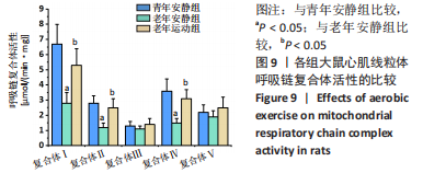
[17] 王增喜,李洁,王悦.高强度间歇训练对大鼠心肌线粒体呼吸链复合体活性的影响[J].中国运动医学杂志,2018,37(4):315-322.
[18] 彭成欢,向常清,张世忠,等.基于线粒体通透性转换孔调控的脓毒症心肌损伤及普拉克索保护机制的研究[J].中国医院药学杂志,2022,42(20):2115-2120.
[19] THAM YK, BERNARDO BC, OOI JY, et al. Pathophysiology of cardiac hypertrophy and heart failure:signaling pathways and novel therapeutic targets. Arch Toxicol. 2015;89(9):1401-1438.
[20] MA ZG, YUAN YP, WU HM, et al. Cardiac fibrosis: new insights into the pathogenesis. Int J Biol Sci. 2018;14(12):1645-1657.
[21] KYSELOVIČ J, LEDDY JJ. Cardiac fibrosis: the beneficial effects of exercise in cardiac fibrosis. Adv Exp Med Biol. 2017;999:257-268.
[22] OLDFIELD CJ, DUHAMEL TA, DHALLA NS. Mechanisms for the transition from physiological to pathological cardiac hypertrophy. Can J Physiol Pharmacol. 2020;98(2):74-84.
[23] GEORGE K, WHYTE GP, GREEN DJ, et al. The endurance athletes heart: acute stress and chronic adaptation. Br J Sports Med. 2012;46(Suppl 1):29-36.
[24] POMATTO L, DAVIES K. Adaptive homeostasis and the free radical theory of ageing. Free Radic Biol Med. 2018;124:420-430.
[25] SHADEL GS, HORVATH TL. Mitochondrial ROS signaling in organismal homeostasis. Cell. 2015;163(3):560-569.
[26] ZOROV DB, JUHASZOVA M, SOLLOTT SJ. Mitochondrial reactive oxygen species (ROS) and ROS-induced ROS release. Physiol Rev. 2014;94(3):909-950.
[27] PFANNER N, WARSCHEID B, WIEDEMANN N. Mitochondrial proteins: from biogenesis to functional networks. Nat Rev Mol Cell Biol. 2019;20(5):267-284.
[28] CAMPOS JC, QUELICONI BB, BOZI L, et al. Exercise reestablishes autophagic flux and mitochondrial quality control in heart failure. Autophagy. 2017;13(8):1304-1317.
[29] BAUER TM, MURPHY E. Role of mitochondrial calcium and the permeability transition pore in regulating cell death. Circ Res. 2020;126(2):280-293.
[30] BERTERO E, MAACK C. Calcium signaling and reactive oxygen species in mitochondria. Circ Res. 2018;122(10):1460-1478.
[31] PELLEGRINO-COPPOLA D. Regulation of the mitochondrial permeability transition pore and its effects on aging. Microb Cell. 2020;7(9):222-233.
[32] THOMAS MM, VIGNA C, BETIK AC, et al. Initiating treadmill training in late middle age offers modest adaptations in Ca2+ handling but enhances oxidative damage in senescent rat skeletal muscle. Am J Physiol Regul Integr Comp Physiol. 2010;298(5):R1269-1278.
[33] PALIKARAS K, LIONAKI E, TAVERNARAKIS N. Coordination of mitophagy and mitochondrial biogenesis during ageing in C. elegans. Nature. 2015;521(7553):525-528.
[34] POPOV LD. Mitochondrial biogenesis: An update. J Cell Mol Med. 2020;24(9):4892-4899.
[35] HALLING JF, PILEGAARD H. PGC-1α-mediated regulation of mitochondrial function and physiological implications. Appl Physiol Nutr Metab. 2020;45(9):927-936.
[36] PICKLES S, VIGIÉ P, YOULE RJ. Mitophagy and quality control mechanisms in mitochondrial maintenance. Curr Biol. 2018;28(4):R170-185.
[37] KIM YA, KIM YS, OH SL, et al. Autophagic response to exercise training in skeletal muscle with age. J Physiol Biochem. 2013;69(4):697-705.
[38] 薄海,李玲,段富强,等.低氧联合运动对大鼠骨骼肌线粒体自噬的影响[J].中国康复医学杂志,2014,29(10):908-912.
[39] ADEBAYO M, SINGH S, SINGH AP, et al. Mitochondrial fusion and fission: the fine-tune balance for cellular homeostasis. FASEB J. 2021;35(6):e21620.
[40] CHAN DC. Mitochondrial dynamics and its involvement in disease. Annu Rev Pathol. 2020;15:235-259.
[41] IKEDA Y, SHIRAKABE A, BRADY C, et al. Molecular mechanisms mediating mitochondrial dynamics and mitophagy and their functional roles in the cardiovascular system. J Mol Cell Cardiol. 2015;78:116-122.
[42] BHANDARI P, SONG M, DORN GW 2ND. Dissociation of mitochondrial from sarcoplasmic reticular stress in Drosophila cardiomyopathy induced by molecularly distinct mitochondrial fusion defects. J Mol Cell Cardiol. 2015;80:71-80.
[43] FANG HY, CHEN CY, CHIOU SH, et al. Overexpression of optic atrophy 1 protein increases cisplatin resistance via inactivation of caspase-dependent apoptosis in lung adenocarcinoma cells. Hum Pathol. 2012;43(1):105-114.
[44] WANG K, LIU CY, ZHANG XJ, et al. miR-361-regulated prohibitin inhibits mitochondrial fission and apoptosis and protects heart from ischemia injury. Cell Death Differ. 2015; 22(6):1058-1068.
[45] JIANG HK, WANG YH, SUN L, et al. Aerobic interval training attenuates mitochondrial dysfunction in rats post-myocardial infarction: roles of mitochondrial network dynamics. Int J Mol Sci. 2014;15(4):5304-5322.
[46] VEERANKI S, GIVVIMANI S, KUNDU S, et al. Moderate intensity exercise prevents diabetic cardiomyopathy associated contractile dysfunction through restoration of mitochondrial function and connexin 43 levels in db/db mice. J Mol Cell Cardiol. 2016;92:163-173.
|