中国组织工程研究 ›› 2023, Vol. 27 ›› Issue (30): 4809-4816.doi: 10.12307/2023.507

• 组织工程骨材料 tissue-engineered bone • 上一篇    下一篇

不同比例羟基磷灰石/β-磷酸三钙涂层支架修复骨缺损

宋美玲1,2,李征宇3,4,艾子政5,李京娜1,2,曾庆丰4,6,韩倩倩7,董谢平1,8,9,10   

  1. 1江西省人民医院骨科,江西省南昌市  330006;2江西中医药大学研究生院,江西省南昌市  330004;3西安市中心医院,陕西省西安市  710003;4点云生物(杭州)有限公司,浙江省杭州市  310018;5南昌市第四医院,江西省南昌市  330104;6迈海材料基因组国际研究院,河北省廊坊市  065500;7中国食品药品检定研究院,北京市  102600;8南昌大学医学院,江西省南昌市  330006;9江西省卫生健康数字骨科重点实验室,江西省南昌市  330006;10南方科技大学医院南方科技大学粤港澳智能与数字外科创新中心,广东省深圳市  518000
  • 收稿日期:2022-06-28 接受日期:2022-08-25 出版日期:2023-10-28 发布日期:2023-04-01
  • 通讯作者: 董谢平,博士生导师,主任医师,江西省人民医院骨科,江西省南昌市 330006;南昌大学医学院,江西省南昌市 330006;江西省卫生健康数字骨科重点实验室,江西省南昌市 330006;南方科技大学医院南方科技大学粤港澳智能与数字外科创新中心,广东省深圳市 518000 韩倩倩,研究员,中国食品药品检定研究院,北京市 102600
  • 作者简介:宋美玲,女,1998年生,江西省瑞金市人,汉族,江西中医药大学在读硕士,主要从事骨组织工程研究。
  • 基金资助:
    国家自然科学基金地区科学基金项目(81860384),项目负责人:董谢平;陕西省重点研发计划一般项目(2019SF-229),项目负责人:李征宇;西安市卫生健康委员会科研项目(2020yb02),项目负责人:李征宇;西安交通大学基本科研业务费自由探索与创新教师类项目(xzy012020052),项目负责人:李征宇

Repair effect of different hydroxyapatite/beta-tricalcium phosphate coated scaffolds on bone defects

Song Meiling1, 2, Li Zhengyu3, 4, Ai Zizheng5, Li Jingna1, 2, Zeng Qingfeng4, 6, Han Qianqian7, Dong Xieping1, 8, 9, 10   

  1. 1Department of Orthopedics, Jiangxi Provincial People’s Hospital, Nanchang 330006, Jiangxi Province, China; 2Graduate School, Jiangxi University of Chinese Medicine, Nanchang 330004, Jiangxi Province, China; 3Xi’an Central Hospital, Xi’an 710003, Shaanxi Province, China; 4Particle Cloud Biotechnology (Hangzhou) Co., Ltd., Hangzhou 310018, Zhejiang Province, China; 5The Fourth Hospital of Nanchang, Nanchang 330104, Jiangxi Province, China; 6MSEA International Institute for Materials Genome, Langfang 065500, Hebei Province, China; 7National Institutes for Food and Drug Control, Beijing 102600, China; 8Medical College, Nanchang University, Nanchang 330006, Jiangxi Province, China; 9Digital Lab of Orthopeadics, Key Laboratory of Health Commission of Jiangxi Province, Nanchang 330006, Jiangxi Province, China; 10Guangdong-Hong Kong-Macao Intelligent and Digital Surgery Innovation Center, Southern University of Science and Technology Hospital, Shenzhen 518000, Guangdong Province, China
  • Received:2022-06-28 Accepted:2022-08-25 Online:2023-10-28 Published:2023-04-01
  • Contact: Dong Xieping, Doctoral supervisor, Chief physician, Department of Orthopedics, Jiangxi Provincial People’s Hospital, Nanchang 330006, Jiangxi Province, China; Medical College, Nanchang University, Nanchang 330006, Jiangxi Province, China; Digital Lab of Orthopeadics, Key Laboratory of Health Commission of Jiangxi Province, Nanchang 330006, Jiangxi Province, China; Guangdong-Hong Kong-Macao Intelligent and Digital Surgery Innovation Center, Southern University of Science and Technology Hospital, Shenzhen 518000, Guangdong Province, China Han Qianqian, Researcher, National Institutes for Food and Drug Control, Beijing 102600, China
  • About author:Song Meiling, Master candidate, Department of Orthopedics, Jiangxi Provincial People’s Hospital, Nanchang 330006, Jiangxi Province, China; Graduate School, Jiangxi University of Chinese Medicine, Nanchang 330004, Jiangxi Province, China
  • Supported by:
    Regional Science Foundation Project of National Natural Science Foundation of China, No. 81860384 (to DXP); General Project of Shaanxi Provincial Key Research & Development Program, No. 2019SF-229 (to LZY); Scientific Research Project of Xi’an Municipal Health Commission, No. 2020yb02 (to LZY); Fundamental Research Fund Free Exploration and Innovation Teacher Project of Xi'an Jiaotong University, No. xzy012020052 (to LZY)

摘要:


文题释义:

临界骨缺损:在动物骨缺损模型中,临界性骨缺损是一重要参数,是指实验动物在自然情况下不能自行愈合的最小尺寸骨缺损。
羟基磷灰石/β-磷酸三钙涂层多孔生物陶瓷支架:生物陶瓷材料具有理化性能稳定、生物相容性优异、耐腐蚀、无毒副作用的特点,被广泛应用于骨组织工程领域。此次实验主要是以羟基磷灰石为基体材料制备3D打印多孔生物陶瓷支架,但羟基磷灰石脆性大、韧性低,为提高支架的生物活性和生物降解性,在多孔生物陶瓷支架表面涂覆不同比例羟基磷灰石/β-磷酸三钙(m∶m)双相磷酸钙陶瓷涂层。

背景:羟基磷灰石/β-磷酸三钙双相磷酸钙陶瓷具有良好的生物相容性与骨传导能力,可以作为涂层材料联合用于超临界骨缺损修复,但羟基磷灰石/β-磷酸三钙涂层的最佳质量比目前尚无明确报道。
目的:在兔桡骨超临界骨缺损部位植入不同比例羟基磷灰石/β-磷酸三钙涂层多孔生物陶瓷支架,评估其修复作用,以期获得最佳的涂层比例。
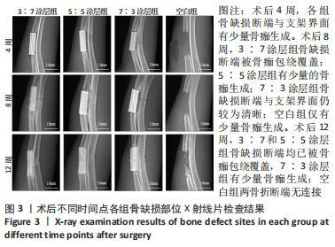
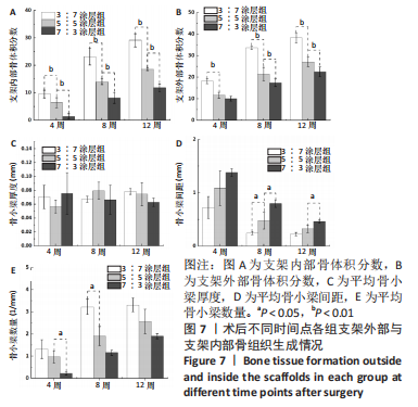
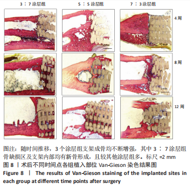
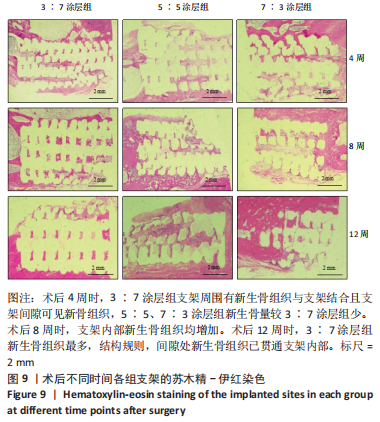
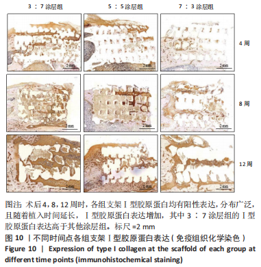
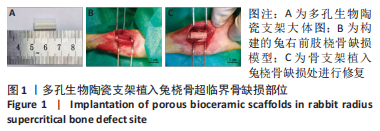
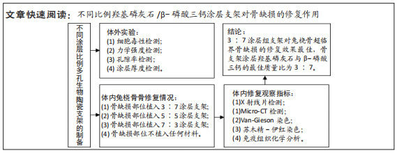
方法:采用3D打印技术制备羟基磷灰石多孔生物陶瓷支架,并在其表面使用不同比例的羟基磷灰石与β-磷酸三钙(二者质量比分别为3∶7,5∶5,7∶3)进行涂层处理,然后对支架的细胞毒性、孔隙率、力学强度、涂层厚度等参数进行测试。在36只新西兰大白兔右前肢建立15.0 mm兔桡骨超临界骨缺损模型,随机分4组处理,每组9只:空白组不植入任何材料,3∶7涂层组、5∶5涂层组、7∶3涂层组分别植入对应质量比例涂层的羟基磷灰石多孔生物陶瓷支架,术后4,8,12周,分别进行X射线片及Micro-CT检查、Van-Gieson染色、苏木精-伊红染色及Ⅰ型胶原蛋白免疫组织化学分析。

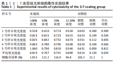
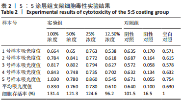
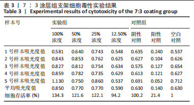
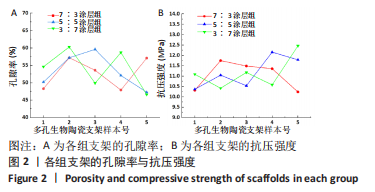
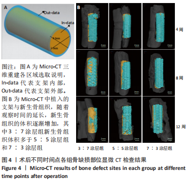
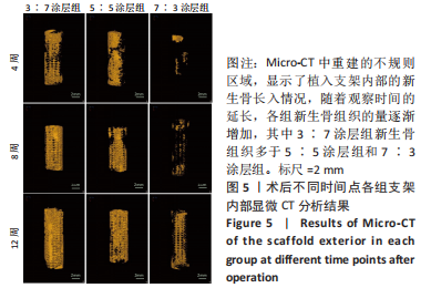
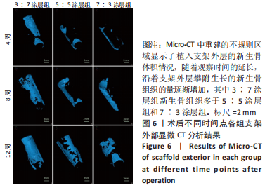
结果与结论:①3∶7、5∶5、7∶3涂层支架的细胞毒性均为0级,涂层厚度为(75.2±0.54) μm,孔隙率分别为(54.02±5.17)%,(53.28± 5.05)%,(52.82±4.55)%,抗压强度分别为(11.15±0.72),(11.18±0.78),(10.24±0.70) MPa;②X射线与Micro-CT检查结果显示,随着时间的增加,各组骨缺损有不同程度的修复,至术后12周时,3∶7涂层组骨缺损修复效果最佳,该组材料内部的骨长入体积与材料外表面的新生骨体积最多;③Van-Gieson染色与苏木精-伊红染色显示,术后12周时,3∶7涂层组骨缺损区及支架内部均有新骨形成,且新生骨组织最多、新生骨结构最为致密;免疫组织化学分析显示,术后12周时,3∶7涂层组Ⅰ型胶原蛋白表达高于其他两组;④结果表明,3∶7涂层组对兔桡骨超临界骨缺损的修复效果最佳,骨支架涂层羟基磷灰石与β-磷酸三钙的最佳质量比为 3∶7。

https://orcid.org/0000-0001-8124-2586(宋美玲)

中国组织工程研究杂志出版内容重点:生物材料;骨生物材料口腔生物材料纳米材料缓释材料材料相容性组织工程

关键词: 骨修复, 骨缺损, 3D生物打印支架, 羟基磷灰石, β-磷酸三钙, 生物陶瓷, 组织工程

Abstract: BACKGROUND: Hydroxyapatite/β-tricalcium phosphate biphasic calcium phosphate ceramics have good biocompatibility and osteoconductivity. They can be used as a coating material for supercritical bone defect repair, but the optimal mass ratio of hydroxyapatite/β-tricalcium phosphate coating has not been reported. 
OBJECTIVE: Porous bioceramic scaffolds with different ratios of hydroxyapatite/β-tricalcium phosphate coating were implanted into the supercritical bone defect site of rabbit radius to assess its repairing effects in order to obtain the best coating ratio.
METHODS: Hydroxyapatite porous bioceramic scaffolds were prepared by 3D printing technology, and coated with different mass ratios of hydroxyapatite/β-tricalcium phosphate (mass ratio: 3:7, 5:5, 7:3, respectively) on their surfaces. Cytotoxicity, porosity, mechanical strength, coating thickness, and other parameters were tested. The 15.0 mm rabbit radial supercritical bone defects were established in the right forelimbs of 36 New Zealand white rabbits. They were randomly divided into four groups (n=9). In the blank group, no material was implanted. In the 3:7 coating group, 5:5 coating group, and 7:3 coating group, hydroxyapatite porous bioceramic scaffolds were implanted with corresponding mass ratio coatings. X-ray, Micro-CT, Van-Gieson staining, hematoxylin-eosin staining, and immunohistochemistry for type I collagen were performed at 4, 8, and 12 weeks after operation. 
RESULTS AND CONCLUSION: (1) The scaffold cytotoxicity of 3:7, 5:5, and 7:3 coating was grade 0. The coating thickness was (75.2±0.54) μm, and the porosity was (54.02±5.17)%, (53.28±5.05)%, and (52.82±4.55)%, respectively. The compressive strength was (11.15±0.72), (11.18±0.78), and (10.24±0.70) MPa, respectively. (2) The X-ray and Micro-CT results showed that with the increase of time, the bone defects in each group were repaired to different degrees. The 3:7 coating group had the best bone defect repair effect at 12 weeks after the operation. The volume of bone ingrowth inside the group of materials and the volume of new bone on the outer surface of the materials were the most. (3) The results of Van-Gieson staining and hematoxylin-eosin staining showed that new bone was formed in the bone defect area and inside the scaffold, and the new bone tissue was the most and the new bone structure was the densest one in the 3:7 coating group at 12 weeks after operation. The results of immunohistochemical analysis showed that the expression of type I collagen in the 3:7 coating group was higher than that in the other two groups at 12 weeks after surgery. (4) The results showed that the 3:7 coating group had the best repair effect on the supercritical bone defects of the rabbits, and the optimal mass ratio of hydroxyapatite/β-tricalcium phosphate was 3:7. 

Key words:  , bone repair, bone defect, 3D bioprinted scaffold, hydroxyapatite, β-tricalcium phosphate, bioceramics, tissue engineering

中图分类号: