[1] 中华医学会骨质疏松和骨矿盐疾病分会.骨质疏松性椎体压缩性骨折诊疗与管理专家共识[J].中华骨质疏松和骨矿盐疾病杂志, 2018,11(5):425-437.
[2] ZHAO G, LIU X, LI F. Balloon kyphoplasty versus percutaneous vertebroplasty for treatment of osteoporotic vertebral compression fractures (OVCFs). Osteoporos Int. 2016;27(9):2823-2834.
[3] GALIBERT P, DERAMOND H, ROSAT P, et al. Preliminary note on the treatment of vertebral angioma by percutaneous acrylic vertebroplasty. Neurochirurgie. 1987;33(2):166-168.
[4] 薛广,杨新明,张瑛. 两种入路行经皮椎体成形治疗胸椎骨质疏松性压缩骨折:骨水泥渗漏及安全性的比较[J]. 中国组织工程研究, 2022,26(28):4514-4518.
[5] MARTINEZ-FERRER A, BLASCO J, CARRASCO JL, et al. Risk factors for the development of vertebral fractures after percutaneous vertebroplasty. J Bone Miner Res. 2013;28(8):1821-1829.
[6] 高剑峰,沈文东,张军强,等.经皮椎体后凸成形术后骨水泥椎间隙渗漏与渗漏椎间盘高度的相关性[J]. 中华骨与关节外科杂志, 2021,14(3):181-185,190.
[7] 黄永恒,刘星,尚显文.骨质疏松性椎体压缩性骨折患者PKP治疗后发生邻近椎体骨折的危险因素分析[J]. 山东医药,2021,61(23): 72-76.
[8] MA X, XING D, MA J, et al. Risk factors for new vertebral compression fractures after percutaneous vertebroplasty: qualitative evidence synthesized from a systematic review. Spine (Phila Pa 1976). 2013; 38(12): E713-722.
[9] 李秋江,房晓敏,王胤斌,等.骨质疏松性椎体压缩性骨折椎体强化术后椎体再骨折的相关因素[J]. 中华骨质疏松和骨矿盐疾病杂志,2021,14(3):252-260.
[10] ZHANG ZL, YANG JS, HAO DJ, et al. Risk Factors for New Vertebral Fracture After Percutaneous Vertebroplasty for Osteoporotic Vertebral Compression Fractures. Clin Interv Aging. 2021;16:1193-1200.
[11] SHIN HK, PARK JH, LEE IG, et al. A study on the relationship between the rate of vertebral body height loss before balloon kyphoplasty and early adjacent vertebral fracture. J Back Musculoskelet Rehabil. 2021;34(4):649-656.
[12] KO BS, CHO KJ, PARK JW. Early Adjacent Vertebral Fractures after Balloon Kyphoplasty for Osteoporotic Vertebral Compression Fractures. Asian Spine J. 2019;13(2):210-215.
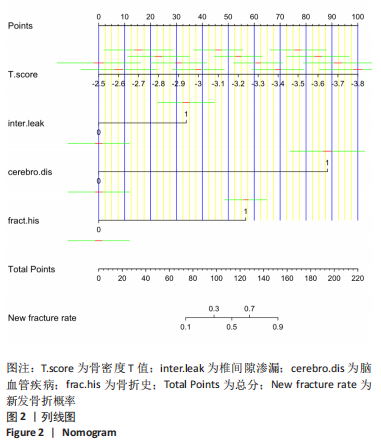
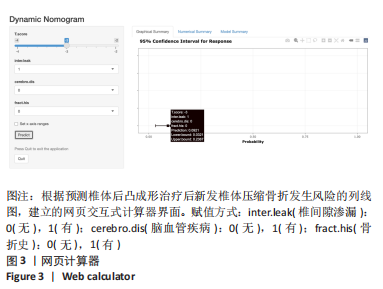
[13] HESS DR. A nomogram for use of non-invasive respiratory strategies in COVID-19. Lancet Digit Health. 2021;3(3): e140-e141.
[14] KARAKOUSIS G, SONDAK VK, ZAGER JS. Overestimation of Risk for Sentinel Lymph Node Metastasis in a Nomogram for T1 Melanomas. J Clin Oncol. 2020;38(27):3234-3235.
[15] 罗伟斌,林勇,孙春喜,等.经皮椎体成形术术后新发椎体骨折特点及危险因素分析[J].中国医药科学,2021,11(10):210-212,224.
[16] 张洋,肖杰,邹伟,等.高原地区椎体成形术后椎体新发骨折的影响因素[J].中国矫形外科杂志,2019,27(6):511-514.
[17] HAAS F, BYRNE NM, REY M. Nomogram for exercise capacity in women. N Engl J Med. 2005;353(21):2301-2303.
[18] 段克友,刘翔宇,熊风,等.绝经后骨质疏松症患者发生骨质疏松性椎体压缩骨折危险因素分析[J]. 山东医药,2021,61(24):34-38.
[19] 夏维波,章振林,林华,等.原发性骨质疏松症诊疗指南(2017)[J].中国骨质疏松杂志,2019,25(3):281-309.
[20] NING L, ZHU J, TIAN S, et al. Correlation Analysis Between Basic Diseases and Subsequent Vertebral Fractures After Percutaneous Kyphoplasty (PKP) for Osteoporotic Vertebral Compression Fractures. Pain Physician. 2021;24(6):E803-E810.
[21] BAYRAM S, AKGÜL T, ADıYAMAN AE, et al. Effect of Sarcopenia on Mortality after Percutaneous Vertebral Augmentation Treatment for Osteoporotic Vertebral Compression Fractures in Elderly Patients: A Retrospective Cohort Study. World Neurosurg. 2020;138:e354-e360.
[22] OSAKI M, OKUDA R, SAEKI Y, et al. Efficiency of coordinator-based osteoporosis intervention in fragility fracture patients: a prospective randomized trial. Osteoporos Int. 2021;32(3):495-503.
[23] BAWA HS, WEICK J, DIRSCHL DR. Anti-Osteoporotic Therapy After Fragility Fracture Lowers Rate of Subsequent Fracture: Analysis of a Large Population Sample. J Bone Joint Surg Am. 2015;97(19): 1555-1562.
[24] 马一阳,杜大江,陈圣宝,等.髋部骨折与心脑血管疾病相关性[J].国际骨科学杂志,2021,42(1):49-53.
[25] 邓晓清,罗玉球,吴彩葵,等.老年人脑卒中后髋部骨折的发生率及相关因素分析[J]. 中华老年医学杂志,2020,39(2):159-163.
[26] WANG HP, SUNG SF, YANG HY, et al. Associations between stroke type, stroke severity, and pre-stroke osteoporosis with the risk of post-stroke fracture: A nationwide population-based study. J Neurol Sci. 2021; 427:117512.
[27] ABDELRASOUL AA, ELSEBAIE NA, GAMALELDIN OA, et al. Imaging of Brain Infarctions: Beyond the Usual Territories. J Comput Assist Tomogr. 2019;43(3):443-451.
[28] RAZEK AAKA, ELSEBAIE NA. Imaging of vascular cognitive impairment. Clin Imaging. 2021;74:45-54.
[29] CALLALY EL, NI CHROININ D, HANNON N, et al. Falls and fractures 2 years after acute stroke: the North Dublin Population Stroke Study. Age Ageing. 2015;44(5):882-886.
[30] BORSCHMANN K, PANG MY, BERNHARDT J, et al. Stepping towards prevention of bone loss after stroke: a systematic review of the skeletal effects of physical activity after stroke. Int J Stroke. 2012;7(4):330-335.
[31] LUTSEY PL, NORBY FL, ENSRUD KE, et al. Association of Anticoagulant Therapy With Risk of Fracture Among Patients With Atrial Fibrillation. JAMA Intern Med. 2020;180(2):245-253.
[32] KLOTZBUECHER CM, ROSS PD, LANDSMAN PB, et al. Patients with prior fractures have an increased risk of future fractures: a summary of the literature and statistical synthesis. J Bone Miner Res. 2000;15(4):721-739.
[33] TAKAHASHI M, NAITOU K, OHISHI T, et al. Comparison of biochemical markers of bone turnover and bone mineral density between hip fracture and vertebral fracture. J Clin Densitom. 2003;6(3):211-218.
[34] INOSE H, KATO T, ICHIMURA S, et al. Risk factors for subsequent vertebral fracture after acute osteoporotic vertebral fractures. Eur Spine J. 2021;30(9):2698-2707.
[35] 张帅,王高举,王清.经皮穿刺椎体后凸成形中骨水泥渗漏入椎管与胸腰椎椎体后壁形态的关系[J]. 中国组织工程研究,2020,24(10): 1477-1483.
[36] 刘东光,周辉,金永明,等.骨质疏松性椎体压缩骨折PVP术后相邻椎体骨折的相关因素分析[J]. 中国脊柱脊髓杂志,2010,20(12): 980-984.
[37] 曹冬子,许正伟,王存良,等.老年骨质疏松性椎体压缩骨折经皮椎体后凸成形术后新发骨折的危险因素分析[J].空军医学杂志, 2018,34(1):41-44. |
