中国组织工程研究 ›› 2022, Vol. 26 ›› Issue (20): 3147-3151.doi: 10.12307/2022.612

• 软骨组织构建 cartilage tissue construction • 上一篇    下一篇

骨关节炎软骨细胞凋亡与组蛋白去乙酰化酶4含量的降低

顾晓东1,李  飞2,车先达2,李鹏翠2   

  1. 1山西白求恩医院,山西省太原市   030032;2山西医科大学第二医院骨与软组织损伤修复山西省重点实验室,山西省太原市   030001
  • 收稿日期:2021-07-03 接受日期:2021-08-19 出版日期:2022-07-18 发布日期:2022-01-18
  • 通讯作者: 李鹏翠,副教授,山西医科大学第二医院,骨与软组织损伤修复山西省重点实验室,山西省太原市 030001
  • 作者简介:顾晓东,男,1984年生,黑龙江省齐齐哈尔市人,汉族,2020年山西医科大学毕业,博士,主治医师,主要从事骨关节炎发病机制及关节软骨损伤修复研究。
  • 基金资助:
    山西省骨关节炎生物学样本资源共享服务平台建设项目(201705D121010),项目负责人:李鹏翠;山西白求恩医院人才引进科研启动金(2021RC016),项目负责人:顾晓东

Relationship between apoptosis of osteoarthritis chondrocytes and reduction of histone deacetylase 4 content

Gu Xiaodong1, Li Fei2, Che Xianda2, Li Pengcui2   

  1. 1Shanxi Bethune Hospital, Taiyuan 030032, Shanxi Province, China; 2Shanxi Key Laboratory of Bone and Soft Tissue Injury Repair, the Second Hospital of Shanxi Medical University, Taiyuan 030001, Shanxi Province, China
  • Received:2021-07-03 Accepted:2021-08-19 Online:2022-07-18 Published:2022-01-18
  • Contact: Li Pengcui, Associate professor, Shanxi Key Laboratory of Bone and Soft Tissue Injury Repair, the Second Hospital of Shanxi Medical University, Taiyuan 030001, Shanxi Province, China
  • About author:Gu Xiaodong, MD, Attending physician, Shanxi Bethune Hospital, Taiyuan 030032, Shanxi Province, China
  • Supported by:
    the Osteoarthritis Biological Sample Resource Sharing Service Platform Project of Shanxi Province, No. 201705D121010 (to LPC); Talent Introduction Research Fund of Shanxi Bethune Hospital, No. 2021RC016 (to GXD)

摘要:

文题释义:
ATF4/CHOP信号通路:内质网应激介导的细胞凋亡主要通过ATF4/CHOP信号通路发挥作用。发生内质网应激时,激活转录因子4的表达上调,上调的激活转录因子4激活其下游CAAT/增强子结合蛋白同源蛋白的表达,从而触发细胞凋亡。
组蛋白去乙酰化酶:是一类催化核心组蛋白N-末端尾部区域赖氨酸残基去乙酰化的蛋白酶,可通过去乙酰化修饰使组蛋白带正电荷,与带负电荷的DNA紧密结合,染色质呈致密卷曲的阻抑结构,进而抑制基因转录,与细胞增殖、细胞肥大分化及细胞凋亡等诸多过程密切相关。组蛋白去乙酰化酶4的分布具有组织特异性,主要分布于脑、肌肉和软骨中。

背景:内质网应激介导的软骨细胞凋亡在骨关节炎的发病中起重要作用,而内质网应激介导的细胞凋亡主要受ATF4/CHOP信号通路调节,但该信号通路在骨关节炎中的作用及其调控仍不清楚。
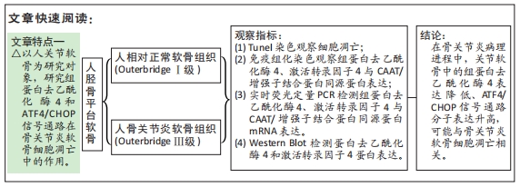
目的:探讨组蛋白去乙酰化酶4和ATF4/CHOP信号通路在骨关节炎软骨细胞凋亡中的作用。
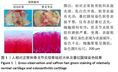
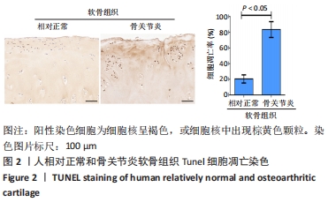
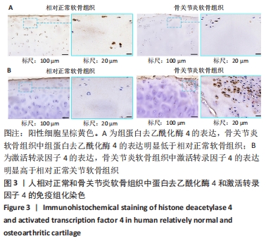
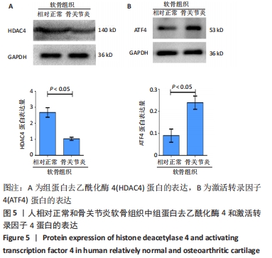
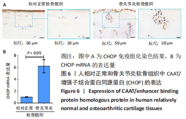
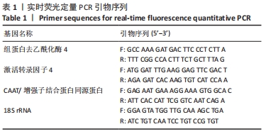
方法:按照Outerbridge分级,将从膝关节置换切取的胫骨平台软骨分为相对正常软骨(OuterbridgeⅠ级)和骨关节炎软骨(Outerbridge Ⅲ级),通过Tunel染色观察软骨细胞凋亡情况,免疫组化染色检测组蛋白去乙酰化酶4、激活转录因子4(ATF4)与CAAT/增强子结合蛋白同源蛋白(CHOP)表达,实时荧光定量PCR检测CAAT/增强子结合蛋白同源蛋白mRNA表达,Western Blot与实时荧光定量PCR检测组蛋白去乙酰化酶4、激活转录因子4的表达。
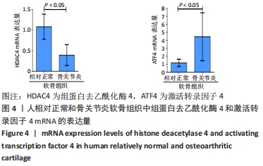
结果与结论:①Tunel染色显示,骨关节炎软骨中软骨细胞的凋亡率高于相对正常软骨[(83.5±10.1)%,(20.5±5.2)%,P < 0.05];②免疫组化染色显示,骨关节炎软骨中激活转录因子4的表达高于相对正常软骨,组蛋白去乙酰化酶4的表达低于相对正常软骨;骨关节炎关节软骨中CAAT/增强子结合蛋白同源蛋白的表达高于相对正常软骨;③骨关节炎软骨中激活转录因子4与CAAT/增强子结合蛋白同源蛋白的mRNA表达均高于相对正常软骨(P < 0.05),组蛋白去乙酰化酶4的mRNA表达低于相对正常软骨(P < 0.05);④骨关节炎软骨中组蛋白去乙酰化酶4的蛋白表达低于相对正常软骨(P < 0.05),激活转录因子4的蛋白表达高于相对正常软骨(P < 0.05);⑤结果表明,在骨关节炎病理进程中,关节软骨中的组蛋白去乙酰化酶4表达降低、ATF4/CHOP信号通路分子表达升高,可能与骨关节炎软骨细胞凋亡相关。
缩略语:CAAT/增强子结合蛋白同源蛋白:CAAT/enhancer-binding protein homologous protein,CHOP

https://orcid.org/0000-0003-1432-6389 (顾晓东) 

中国组织工程研究杂志出版内容重点:组织构建;骨细胞;软骨细胞;细胞培养;成纤维细胞;血管内皮细胞;骨质疏松;组织工程

关键词: 骨关节炎, 软骨细胞, 组蛋白去乙酰化酶4, 激活转录因子4, CAAT/增强子结合蛋白同源蛋白, 内质网应激, 凋亡

Abstract: BACKGROUND: Endoplasmic reticulum stress-mediated chondrocyte apoptosis plays an important role in the pathogenesis of osteoarthritis, which is mainly regulated by the activated transcription factor 4 (ATF4)/CAAT/enhancer-binding protein homologous protein (CHOP) signaling pathway. However, the mechanism of this signaling pathway in osteoarthritis remains unclear.
OBJECTIVE: To investigate the mechanism of histone deacetylase 4 and ATF4/CHOP signaling pathway in osteoarthritis chondrocytes apoptosis.
METHODS: According to Outerbridge classification, the articular cartilage of the tibial plateau cartilage obtained during knee joint replacement was divided into relatively normal cartilage (Outerbridge I) and osteoarthritis cartilage (Outerbridge III). Apoptosis of chondrocytes was observed by TUNEL staining. Immunohistochemistry staining was used to detect the expression of histone deacetylase 4, ATF4, and CHOP. Real-time fluorescence quantitative PCR was used to detect the mRNA expression of CHOP, while western blot and real-time fluorescence quantitative PCR were used to detect the protein expression of histone deacetylase 4 and ATF4.
RESULTS AND CONCLUSION: TUNEL staining results showed that the apoptosis rate of chondrocytes in osteoarthritic cartilage was higher than that in relatively normal cartilage [(83.5±10.1)% vs. (20.5±5.2)%, P < 0.05]. Immunohistochemical staining results showed that the expression of ATF4 in osteoarthritic cartilage was significantly higher than that in relatively normal cartilage, and the expression of histone deacetylase 4 in osteoarthritic cartilage was lower than that in relatively normal cartilage. The expression of CHOP in osteoarthritis cartilage was higher than that in relatively normal cartilage. The mRNA expression of ATF4 and CHOP in osteoarthritic cartilage were higher than that in relative normal cartilage (P < 0.05), and the mRNA expression of histone deacetylase 4 in osteoarthritic cartilage was lower than that in relative normal cartilage (P < 0.05). The protein expression of histone deacetylase 4 in osteoarthritic cartilage was lower than that in relatively normal cartilage (P < 0.05), and the protein expression of ATF4 in osteoarthritic cartilage was higher than that in relatively normal cartilage (P < 0.05). Therefore, in the pathological process of osteoarthritis, the downregulated expression of histone deacetylase 4 and upregulated expression of ATF4/CHOP signal pathway molecules may be related to chondrocyte apoptosis in osteoarthritis.

Key words: osteoarthritis, chondrocyte, histone deacetylase 4, activating transcription factor 4, CAAT/enhancer binding protein homologous protein, endoplasmic reticulum stress, apoptosis

中图分类号: