[1] WANG HW, LAI EH, YANG CN, et al. Intracanal Metformin Promotes Healing of Apical Periodontitis via Suppressing Inducible Nitric Oxide Synthase Expression and Monocyte Recruitment.J Endod. 2020;46:65-73.
[2] RÔÇAS I, SIQUEIRA J. Frequency and levels of candidate endodontic pathogens in acute apical abscesses as compared to asymptomatic apical periodontitis. PLoS ONE.2018;13:e0190469.
[3] Siqueira JF Jr, Antunes HS, Pérez AR, et al. The Apical Root Canal System of Teeth with Posttreatment Apical Periodontitis: Correlating Microbiologic, Tomographic, and Histopathologic Findings. J Endod. 2020; 46(9):1195-1203.
[4] NIKOLIC N, JAKOVLJEVIC A, CARKIC J, et al. Notch Signaling Pathway in Apical Periodontitis: Correlation with Bone Resorption Regulators and Proinflammatory Cytokines. J Endod. 2019;45:123-128.
[5] TAIRA TM, LIMA V, PRADO DS, et al. NLRP12 Attenuates Inflammatory Bone Loss in Experimental Apical Periodontitis. J Dent Res. 2019;98:476-484.
[6] WANG L, ZHANG H, DONG M, et al. Role of the Btk-PLCγ2 Signaling Pathway in the Bone Destruction of Apical Periodontitis. Mediators Inflamm. 2019;2019:8767529.
[7] PASPARAKIS M, VANDENABEELE P. Necroptosis and its role in inflammation. Nature. 2015;517:311-320.
[8] WEINLICH R, OBERST A, BEERE HM, et al. Necroptosis in development, inflammation and disease. Nat Rev Mol Cell Biol. 2017;18(2):127-136.
[9] KHOURY MK, GUPTA K, FRANCO SR, et al. Necroptosis in the Pathophysiology of Disease. Am J Pathol. 2020;190(2):272-285.
[10] WU Y, SUN H, YANG B,et al. 5-Lipoxygenase Knockout Aggravated Apical Periodontitis in a Murine Model. J Dent Res. 2018;97:442-450.
[11] TAKAHAMA A, RÔÇAS I, FAUSTINO I, et al. Association between bacteria occurring in the apical canal system and expression of bone-resorbing mediators and matrix metalloproteinases in apical periodontitis. Int Endod J. 2018;51(7):738-746.
[12] MÁRTON I, KISS C. Overlapping protective and destructive regulatory pathways in apical periodontitis. J Endod. 2014;40:155-163.
[13] TIBÚRCIO-MACHADO CS, MICHELON C, ZANATTA FB, et al. The global prevalence of apical periodontitis: a systematic review and meta-analysis. Int Endod J. 2020 Dec 30. doi: 10.1111/iej.13467.
[14] Pirani C, Iacono F, Gatto MR, et al. Outcome of secondary root canal treatment filled with Thermafil: a 5-year follow-up of retrospective cohort study. Clin Oral Investig. 2018;22(3):1363-1373.
[15] Xu R, Guo D, Zhou X, et al. Disturbed bone remodelling activity varies in different stages of experimental, gradually progressive apical periodontitis in rats. Int J Oral Sci. 2019;11(3):27.
[16] Jun-Long H, Yi L, Bao-Lian Z, et al. Necroptosis Signaling Pathways in Stroke: From Mechanisms to Therapies. Curr Neuropharmacol. 2018; 16(9):1327-1339.
[17] Wang L, Du F, Wang X. TNF-alpha induces two distinct caspase-8 activation pathways. Cell. 2008;133:693-703.
[18] Ofengeim D, Yuan J. Regulation of RIP1 kinase signalling at the crossroads of inflammation and cell death. Nat Rev Mol Cell Biol. 2013; 14(11):727-736.
[19] Shan B, Pan H, Najafov A, et al. Necroptosis in development and diseases.Genes Dev. 2018;32(5-6):327-340.
[20] De Rossi A, Lucisano MP, De Rossi M, et al. Effect of intercellular adhesion molecule 1 deficiency on the development of apical periodontitis. Int Endod J.2020;53(3):354-365.
[21] Pierdomenico M, Negroni A, Stronati L, et al. Necroptosis is active in children with inflammatory bowel disease and contributes to heighten intestinal inflammation.Am J Gastroenterol. 2014;109(2):279-287.
[22] Lee JM, Yoshida M, Kim MS, et al. Involvement of Alveolar Epithelial Cell Necroptosis in Idiopathic Pulmonary Fibrosis Pathogenesis. Am J Respir Cell Mol Biol.2018;59(2):215-224.
[23] Ke X, Lei L, Li H, et al. Manipulation of necroptosis by Porphyromonas gingivalis in periodontitis development. Mol Immunol. 2016;77:8-13.
[24] Brennan CA, Garrett WS. Fusobacterium nucleatum-symbiont, opportunist and oncobacterium. Nat Rev Microbiol. 2019;17(3):156-166.
[25] Redlich K, Smolen JS. Inflammatory bone loss: pathogenesis and therapeutic intervention. Nat Rev Drug Discov. 2012;11(3):234-250.
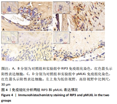
|