[1] XIA WH, LI J, SU C, et al. Physical exercise attenuates age-associated reduction in endothelium-reparative capacity of endothelial progenitor cells by increasing CXCR4/JAK-2 signaling in healthy men. Aging Cell. 2012;11(1):111-119.
[2] ASAHARA T, MUROHARA T, SULLIVAN A, et al. Isolation of putative progenitor endothelial cells for angiogenesis. Science. 1997;275(5302): 964-967.
[3] CHILLÀ A, MARGHERI F, BIAGIONI A, et al. Mature and progenitor endothelial cells perform angiogenesis also under protease inhibition: the amoeboid angiogenesis. J Exp Clin Cancer Res. 2018;37(1):74.
[4] XUE C, HUANG Q, ZHANG T, et al. Matrix stiffness regulates arteriovenous differentiation of endothelial progenitor cells during vasculogenesis in nude mice. Cell Prolif. 2019;52(2):e12557.
[5] HU Z, WANG H, FAN G, et al. Danhong injection mobilizes endothelial progenitor cells to repair vascular endothelium injury via upregulating the expression of Akt, eNOS and MMP-9. Phytomedicine. 2019;61: 152850.
[6] FANG J, GUO Y, TAN S, et al. Autologous Endothelial Progenitor Cells Transplantation for Acute Ischemic Stroke: A 4-Year Follow-Up Study. Stem Cells Transl Med. 2019;8(1):14-21.
[7] 秦臻,韦正新,宰青青,等.黄精降低活性氧水平促进衰老内皮祖细胞功能的研究[J].中国药理学通报,2019,35(1):123-127.
[8] 朱光旭,黄岚,武晓静,等.不同年龄段大鼠血清影响内皮祖细胞向成熟内皮诱导分化[J].中国动脉硬化杂志,2007,15(9): 647-652.
[9] OU HL, SCHUMACHER B. DNA damage responses and p53 in the aging process. Blood. 2018;131(5):488-495.
[10] 秦臻,韦正新,许键炜.黄精对衰老大鼠内皮祖细胞 DNA 损伤检测点ATM/ATR 通路的影响[J].中药新药与临床药理,2019,30(5):529-534.
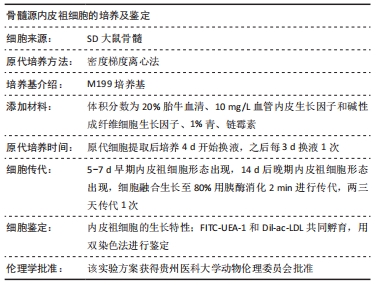
[11] NGANGUEM NZALLE YRANNEY BRICE,林慕之,毛善永,等.大鼠骨髓源内皮祖细胞的分离与鉴定[J].贵州医科大学学报,2020,45(3): 265-269.
[12] 李丽玲,计晓娟,余更生,等.1种新来源的内皮祖细胞提取分离方法[J].免疫学杂志,2019,35(3):269-276.
[13] 罗伟,李翔翮,杨先腾,等.小鼠骨髓源性内皮祖细胞的分离培养与鉴定[J].中华创伤骨科杂志,2018,20(6):523-528.
[14] 秦臻,黄水清.当归补血汤含药血清对内皮祖细胞功能及其 PI3K/Akt 通路影响的研究[J].中国药理学通报,2013,29(9):1320-1323.
[15] 胡洁,陈庆伟,贺无恙.增龄与动脉粥样硬化性心血管疾病临床类型的相关性分析[J].临床心血管病杂志,2018,34(11):1081-1085.
[16] 饶璇,李元建.内皮细胞损伤与修复的研究进展[J].中国动脉硬化杂志,2017,25(5):531-535.
[17] FAZEL S, CIMINI M, CHEN L, et al. Cardioprotective c-kit+ cells are from the bone marrow and regulate the myocardial balance of angiogenic cytokines. J Clin Invest. 2006;116(7):1865-1877.
[18] QIAO J, QI K, CHU P, et al. Infusion of endothelial progenitor cells ameliorates liver injury in mice after haematopoietic stem cell transplantation. Liver Int. 2015;35(12):2611-2620.
[19] 宰青青,秦臻,叶兰.黄精对自然衰老大鼠内皮祖细胞功能及端粒酶活性的影响[J].中国中西医结合杂志,2016,36(12):1480-1485.
[20] NAKAMURA AJ, CHIANG YJ, HATHCOCK KS, et al. Both telomeric and non-telomeric DNA damage are determinants of mammalian cellular senescence. Epigenetics Chromatin. 2008;1(1):6.
[21] 李文瑞,刘新光,周中军. DNA 损伤与细胞衰老关系的研究进展[J].国际检验医学杂志,2010,31(6):576-578.
[22] MAYNARD S, FANG EF, SCHEIBYE-KNUDSEN M, et al. DNA Damage, DNA Repair, Aging, and Neurodegeneration. Cold Spring Harb Perspect Med. 2015;5(10):a025130.
[23] MARÉCHAL A, ZOU L. DNA damage sensing by the ATM and ATR kinases. Cold Spring Harb Perspect Biol. 2013;5(9):a012716.
[24] 乔田奎,陈伟,袁素娟.ATM 蛋白激酶依赖性信号转导通路研究进展[J].复旦学报(医学版),2012,39(4):433-437.
[25] FALCK J, COATES J, JACKSON SP. Conserved modes of recruitment of ATM, ATR and DNA-PKcs to sites of DNA damage. Nature. 2005; 434(7033):605-611.
[26] SANCAR A, LINDSEY-BOLTZ LA, UNSAL-KAÇMAZ K, et al. Molecular mechanisms of mammalian DNA repair and the DNA damage checkpoints. Annu Rev Biochem. 2004;73:39-85.
[27] REAPER PM, GRIFFITHS MR, LONG JM, et al. Selective killing of ATM- or p53-deficient cancer cells through inhibition of ATR. Nat Chem Biol. 2011;7(7):428-430.
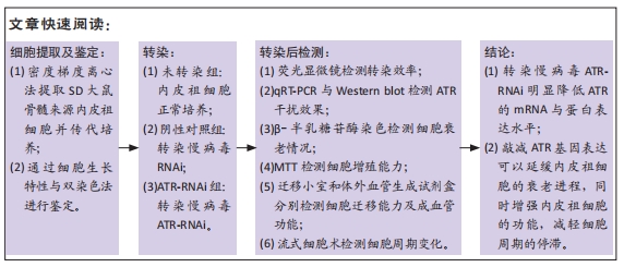
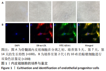
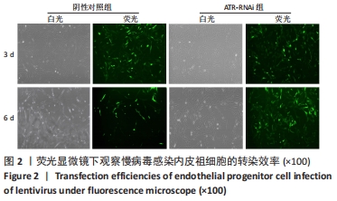
|