[1] GRAHAM SM, LEONIDOU A, ASLAM-PERVEZ N, et al. Biological therapy of bone defects: the immunology of bone allo-transplantation. Expert Opin Biol Ther. 2010;10(6):885-901.
[2] AHMED GA, ISHAQUE B, RICKERT M, et al. Allogeneic bone transplantation in hip revision surgery : Indications and potential for reconstruction. Orthopade. 2018;47(1):52-66.
[3] CHU W. Treatment of nonunion with autologous bone transplantation combined with platelet-rich plasma and extracorporeal shock wave. Zhongguo Gu Shang. 2019;32(5):434-439.
[4] 余幸鸽,林开利.基于天然水凝胶的生物材料在骨组织工程中的应用[J].中国生物工程杂志,2020,40(5):69-77.
[5] HOLZAPFEL BM, RUDERT M, HUTMACHER DW. Scaffold-based Bone Tissue Engineering. Orthopade. 2017;46(8):701-710.
[6] ZHOU X, SHI G, FAN B, et al. Polycaprolactone electrospun fiber scaffold loaded with iPSCs-NSCs and ASCs as a novel tissue engineering scaffold for the treatment of spinal cord injury. Int J Nanomedicine. 2018;13:6265-6277.
[7] 武小童,何儿,刘来俊,等.基于聚己内酯纤维的组织工程支架研究进展[J].中国生物医学工程学报,2020,39(5):611-620.
[8] TEOH SH, GOH BT, LIM J. Three-Dimensional Printed Polycaprolactone Scaffolds for Bone Regeneration Success and Future Perspective. Tissue Eng Part A. 2019;25(13-14):931-935.
[9] ALEMÁN-DOMÍNGUEZ ME, GIUSTO E, ORTEGA Z, et al. Three-dimensional printed polycaprolactone-microcrystalline cellulose scaffolds. J Biomed Mater Res B Appl Biomater. 2019;107(3):521-528.
[10] DAAS I, BADR S, OSMAN E. Comparison between Fluoride and Nano-hydroxyapatite in Remineralizing Initial Enamel Lesion: An in vitro Study. J Contemp Dent Pract. 2018;19(3):306-312.
[11] GIZER M, KÖSE S, KARAOSMANOGLU B, et al. The Effect of Boron-Containing Nano-Hydroxyapatite on Bone Cells.Biol Trace Elem Res. 2020;193(2):364-376.
[12] 胡超然,邱冰,周祝兴,等.3D打印聚己内酯/纳米羟基磷灰石复合支架与骨髓间充质干细胞的体外生物相容性[J].中国组织工程研究,2020,24(4):589-595.
[13] NAUDOT M, GARCIA GARCIA A, JANKOVSKY N, et al. The combination of a poly-caprolactone/nano-hydroxyapatite honeycomb scaffold and mesenchymal stem cells promotes bone regeneration in rat calvarial defects. J Tissue Eng Regen Med. 2020;14(11):1570-1580.
[14] ROBERTS EAH, CARTWRIGHT RA, OLDSCHOOL M. The phenolic substances of manufactured tea. I. —Fractionation and paper chromatography of water‐soluble substances. J Sci Food Agric. 1957; 8(2):72-80.
[15] 尚希福,路玉峰,张文志,等.茶黄素对人骨髓基质干细胞的成骨诱导作用[J].山东医药,2010,50(19):7-9.
[16] 赵传勇,丁艳芳,张文志,等.骨髓间充质干细胞联合茶黄素修复激素性股骨头坏死[J].中国组织工程研究,2015,19(32):5210-5214.
[17] Oka Y, Iwai S, Amano H, et al. Tea Polyphenols Inhibit Rat Osteoclast Formation and Differentiation. J Pharmacol Sci. 2012;118(1):55-64.
[18] 辛红美,许洁,汪长东.淫羊藿苷促进MC3T3-E1成骨分化通过Hedgehog信号通路[J].中国药理学通报,2020,36(5):616-620.
[19] 向声燚,焦志伟,马昊鹏,等.聚己内酯/纳米羟基磷灰石复合材料的3D打印及性能[J].工程塑料应用,2018,46(8):122-125,130.
[20] JIN Y, ZHANG W, LIU Y, et al. rhPDGF-BB via ERK pathway osteogenesis and adipogenesis balancing in ADSCs for critical-sized calvarial defect repair. Tissue Eng Part A. 2014;20(23-24):3303-3313.
[21] INODA H, YAMAMOTO G, HATTORI T. Histological investigation of osteoinductive properties of rh-BMP2 in a rat calvarial bone defect model. J Craniomaxillofac Surg. 2004;32(6):365-369.
[22] 李树源,周琦石,周宏亮,等.强骨胶囊辅助诱导膜技术治疗感染性骨髓炎并骨缺损临床观察[J].山东医药,2019,59(19):73-76.
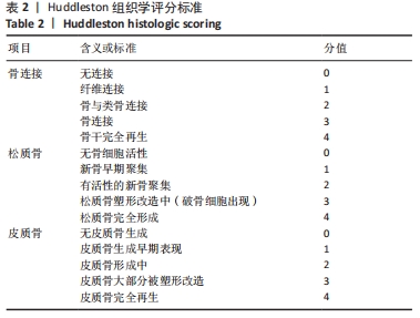
[23] HUDDLESTON PM, STECKELBERG JM, HANSSEN AD, et al. Ciprofloxacin inhibition of experimental fracture healing. J Bone Joint Surg Am. 2000; 82(2):161-173.
[24] FENNEMA EM, TCHANG LAH, YUAN H, et al. Ectopic bone formation by aggregated mesenchymal stem cells from bone marrow and adipose tissue: A comparative study. J Tissue Eng Regen Med. 2018;12(1): e150-e158.
[25] 董晨露.骨髓间充质干细胞成骨分化的研究与进展[J].中国骨伤杂志,2019,32(3):288-292.
[26] NISHIKAWA K, IWAMOTO Y, KOBAYASHI Y, et al. DNA methyltransferase 3a regulates osteoclast differentiation by coupling to an S-adenosylmethionine-producing metabolic pathway. Nat Med. 2015; 21(3):281-287.
[27] CHAI S, WAN L, WANG JL, et al. Gushukang inhibits osteocyte apoptosis and enhances BMP-2/Smads signaling pathway in ovariectomized rats. Phytomedicine. 2019;64:153063.
[28] FENG C, XIAO L, YU JC, et al. Simvastatin promotes osteogenic differentiation of mesenchymal stem cells in rat model of osteoporosis through BMP-2/Smads signaling pathway. Eur Rev Med Pharmacol Sci. 2020;24(1):434-443.
[29] MOON SH, KIM I, KIM SH. Mollugin enhances the osteogenic action of BMP-2 via the p38-Smad signaling pathway. Arch Pharm Res. 2017; 40(11):1328-1335.
[30] CHUNG YH, HUANG YH, CHU TH, et al. BMP-2 restoration aids in recovery from liver fibrosis by attenuating TGF-beta1 signaling. Lab Invest. 2018;98(8):999-1013.
[31] REN C, GONG W, LI F, et al. Pilose antler aqueous extract promotes the proliferation and osteogenic differentiation of bone marrow mesenchymal stem cells by stimulating the BMP-2/Smad1, 5/Runx2 signaling pathway. Chin J Nat Med. 2019;17(10):756-767.
[32] YANG G, YUAN G, LI X, et al. BMP-2 induction of Dlx3 expression is mediated by p38/Smad5 signaling pathway in osteoblastic MC3T3-E1 cells. J Cell Physiol. 2014;229(7):943-954.
[33] YODTHONG T, KEDJARUNE-LEGGAT U, SMYTHE C, etal.l-Quebrachitol Promotes the Proliferation, Differentiation, and Mineralization of MC3T3-E1 Cells: Involvement of the BMP-2/Runx2/MAPK/Wnt/beta-Catenin Signaling Pathway. Molecules. 2018;23(12):3086.
[34] MOON SH, KIM I, KIM SH. Mollugin enhances the osteogenic action of BMP-2 via the p38-Smad signaling pathway. Arch Pharm Res. 2017; 40(11):1328-1335.
[35] 金光辉,孙晓飞,夏琰,等.选择性激光烧结法构建纳米羟基磷灰石与聚己内酯复合材料人工骨支架[J].第二军医大学学报,2015, 36(12): 1289-1294.
[36] 金光辉,张馨雯,孙晓飞,等.组织工程化纳米羟基磷灰石/聚己内酯人工骨支架修复兔桡骨大段骨缺损的实验研究[J].中华损伤与修复杂志(电子版),2015(1):43-49.
[37] QIN T, LI X, LONG H, et al. Bioactive Tetracalcium Phosphate Scaffolds Fabricated by Selective Laser Sintering for Bone Regeneration Applications. Materials (Basel). 2020;13(10):2268.
|