中国组织工程研究 ›› 2022, Vol. 26 ›› Issue (16): 2487-2491.doi: 10.12307/2022.245

• 材料生物相容性 material biocompatibility • 上一篇    下一篇

可生物降解植物多糖止血微球的制备及性能

杨  雪1,王宝群1,姜晓文1,邹圣灿1,明津法2,林莎莎1   

  1. 1青岛琛蓝海洋生物工程有限公司,山东省青岛市  266100;2青岛大学纺织服装学院,山东省青岛市  266000
  • 收稿日期:2020-12-17 修回日期:2020-12-22 接受日期:2021-01-26 出版日期:2022-06-08 发布日期:2021-10-29
  • 通讯作者: 林莎莎,硕士,青岛琛蓝海洋生物工程有限公司,山东省青岛市 266100
  • 作者简介:杨雪,女,1990年生,山东省威海市人,硕士,主要从事生物医用材料相关研究
  • 基金资助:
    省部共建生物多糖纤维成形与生态纺织国家重点实验室(青岛大学)自主课题(重点课题)(RZ2000003348),项目负责人:明津法

Preparation and properties of biodegradable plant polysaccharide hemostatic microspheres

Yang Xue1, Wang Baoqun1, Jiang Xiaowen1, Zou Shengcan1, Ming Jinfa2, Lin Shasha1   

  1. 1Qingdao Chenland Marine Biotechnology Co., Ltd., Qingdao 266100, Shandong Province, China; 2College of Textiles and Clothing, Qingdao University, Qingdao 266000, Shandong Province, China
  • Received:2020-12-17 Revised:2020-12-22 Accepted:2021-01-26 Online:2022-06-08 Published:2021-10-29
  • Contact: Lin Shasha, Master, Qingdao Chenland Marine Biotechnology Co., Ltd., Qingdao 266100, Shandong Province, China
  • About author:Yang Xue, Master, Qingdao Chenland Marine Biotechnology Co., Ltd., Qingdao 266100, Shandong Province, China
  • Supported by:
    An Independent Project of the State Key Laboratory of Biopolysaccharide Fiber Forming and Eco-textile (Qingdao University) Jointly Established by the Province and the Ministry, No. RZ2000003348 (to MJF)

摘要:

文题释义:

微孔多糖止血微球:为一种新型止血材料,具有良好的生物兼容性,无毒副作用,能直接作用于手术创面迅速止血。
淀粉糊化:是将淀粉混合于水中并加热,达到一定温度后淀粉粒溶胀、崩溃,形成黏稠均匀的透明糊溶液。

背景:止血是临床治疗的重要部分,快速有效的止血是手术患者生命安全的必要保障。
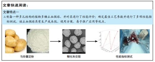
目的:制备一种具有多孔结构的植物多糖止血微球,评价其性能。
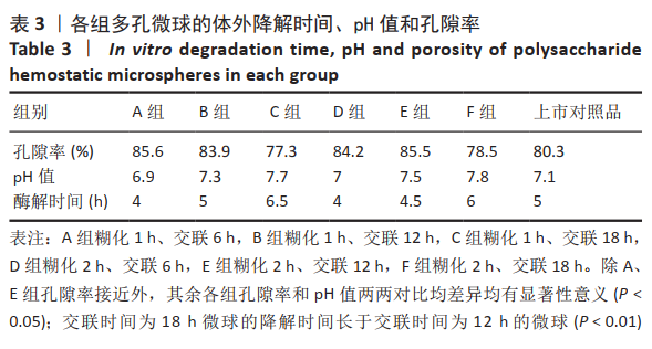
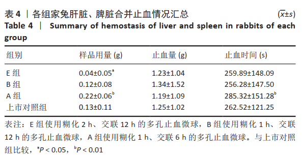
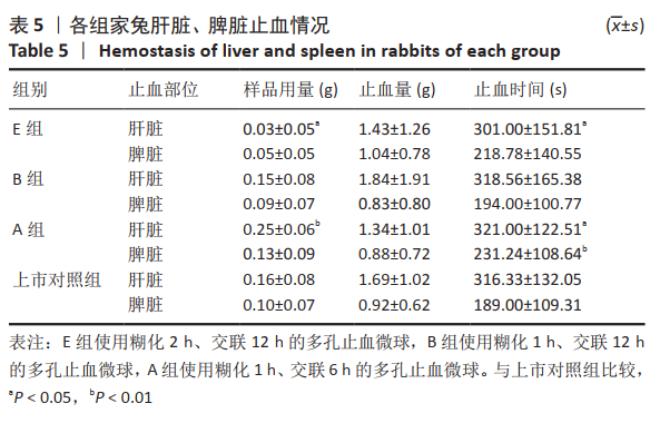
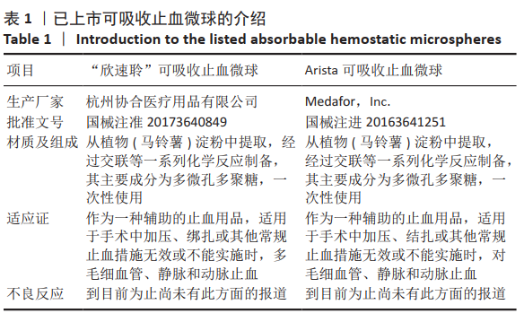
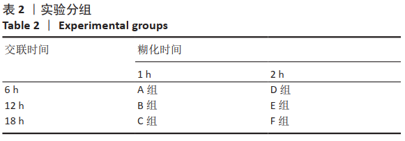
方法:以马铃薯淀粉为原料,通过糊化、乳化交联、清洗、干燥技术制备多孔止血微球,其中通过改进淀粉的糊化和交联工艺,以提高止血微球的止血性能,设置分组:A组糊化1 h、交联6 h,B组糊化1 h、交联12 h,C组糊化1 h、交联18 h,D组糊化2 h、交联6 h,E组糊化2 h、交联12 h,F组糊化2 h、交联18 h,以上市的可吸收止血微球为对照,检测各组微球的微观形貌、pH值、孔隙率与体外降解情况。根据上述实验结果,选择适宜的微球用于新西兰兔肝脏与脾脏的止血,对比样品用量、止血量与止血时间。

结果与结论:①扫描电镜显示,B组与E组形成了直径为50-200 μm结构稳定的多孔微球,成球形态及孔隙率优于对照组;除C组和E组外,其他样品的pH值均在6.5-7.5之间,符合pH值要求;相同质量的样品,交联时间增长降解时间明显增长(P < 0.01)。根据前期形态结构、孔隙率、pH值和体外降解的检测,选择A、B、E组和对照组进行止血实验。②在相同的止血效果下,与对照组比较,E组微球的止血时间更短、用量更少,B组止血效果与对照组比较无明显差异。③结果表明,在60 ℃下、糊化2 h、乳化45 min、交联12 h后制备的多孔淀粉微球,止血效果最佳且其降解时间也相对较短,其性能优于上市对照品。

https://orcid.org/0000-0002-6294-3368 (杨雪) 

中国组织工程研究杂志出版内容重点:生物材料;骨生物材料口腔生物材料纳米材料缓释材料材料相容性;组织工程

关键词: 多孔微球, 结构分析, 制备方法, 糊化, 交联, 体外降解, 止血,

Abstract: BACKGROUND: Hemostasis is an important part of clinical treatment. Fast and effective hemostasis is necessary to ensure the safety of surgical patients.
OBJECTIVE: To prepare a kind of plant polysaccharide hemostatic microsphere with porous structure and its performance evaluation. 
METHODS: Porous hemostatic microspheres were prepared from potato starch by gelatinization, emulsification crosslinking, cleaning, and drying technique. The hemostatic performance of the hemostatic microspheres was improved by improving the gelatinization and crosslinking processes of the starches, setting the grouping: group A (gelatinization for 1 hour, crosslinking for 6 hours, group B (gelatinization for 1 hour, crosslinking for 12 hours), group C (gelatinization for        1 hour, crosslinking for 18 hours), group D (gelatinization for 2 hours, crosslinking for 6 hours), group E (gelatinization for 2 hours, crosslinking for 12 hours), and  group F (gelatinization for 2 hours and crosslinking for 18 hours). The listed absorbable hemostatic microspheres were used as controls to examine the microtopography, pH, porosity and in vitro degradation of microspheres in each group. According to the above experimental results, suitable microspheres were selected for hemostasis of liver and spleen in New Zealand rabbits, comparing the sample dosage, hemostasis amount with hemostasis time.
RESULTS AND CONCLUSION: (1) Scanning electron microscope showed that group B and group E formed porous microspheres with a stable structure of 50-200 μm in diameter, and the spherical morphology and porosity were better than those of the control group. Except for groups C and E, the pH values of the other samples were in the range of 6.5-7.5, which met the pH requirements. The same quality of samples, the cross-linking time growth and degradation time increased significantly (P < 0.01). Based on the examination of morphological structure, porosity, pH and in vitro degradation in the previous stage, groups A, B, E and the control group were selected for hemostasis experiments. (2) Under the same hemostatic effect, the hemostatic time of microspheres in group E was shorter and less used compared with that in the control group, and the hemostatic effect in group B was not significantly different from that in the control group. (3) The results showed that the porous starch microspheres prepared after gelatinization at 60 °C for 2 hours, emulsification for 45 minutes, and cross-linking for 12 hours had the best hemostatic effect and its degradation time was also relatively short, and its performance was superior to that of listed products. 

Key words: porous microspheres, structural analysis, preparation method, gelatinization, crosslinking, in vitro degradation, hemostasis, rabbit

中图分类号: