Chinese Journal of Tissue Engineering Research ›› 2023, Vol. 27 ›› Issue (34): 5441-5447.doi: 10.12307/2023.803
Previous Articles Next Articles
Biological properties of nano-hydroxyapatite-zinc oxide composite scaffolds and their effects on the behavior of MC3T3-E1 osteoblasts
Liu Zixuan1, Li Yan1, 2, Ji Lin1, Xia Delin1, 3
- 1Department of Oral and Maxillofacial Surgery, Affiliated Stomatological Hospital of Southwest Medical University, Luzhou 646000, Sichuan Province, China; 2Taizhou Polytechnic College, Bone Tissue Engineering Research Center of Taizhou, Taizhou 225300, Jiangsu Province, China; 3Department of Plastic and Maxillofacial Surgery, Second Affiliated Hospital of Chongqing Medical University, Chongqing 400010, China
-
Received:2022-09-22Accepted:2022-10-31Online:2023-12-08Published:2023-04-20 -
Contact:Xia Delin, MD, Professor, Chief physician, Department of Oral and Maxillofacial Surgery, Affiliated Stomatological Hospital of Southwest Medical University, Luzhou 646000, Sichuan Province, China; Department of Plastic and Maxillofacial Surgery, Second Affiliated Hospital of Chongqing Medical University, Chongqing 400010, China -
About author:Liu Zixuan, Master candidate, Department of Oral and Maxillofacial Surgery, Affiliated Stomatological Hospital of Southwest Medical University, Luzhou 646000, Sichuan Province, China -
Supported by:Natural Science Project of Colleges and Universities of Jiangsu Province, No. 21KJB180001 (to LY); Jiangsu “Qinglan Project” Outstanding Young Backbone Teachers Project (2021) (to LY)
CLC Number:
Cite this article
Liu Zixuan, Li Yan, Ji Lin, Xia Delin. Biological properties of nano-hydroxyapatite-zinc oxide composite scaffolds and their effects on the behavior of MC3T3-E1 osteoblasts[J]. Chinese Journal of Tissue Engineering Research, 2023, 27(34): 5441-5447.
share this article
Add to citation manager EndNote|Reference Manager|ProCite|BibTeX|RefWorks
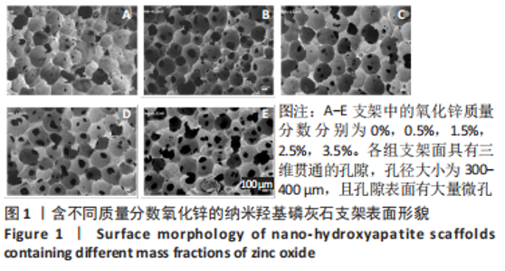
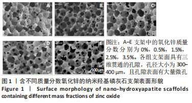
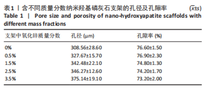
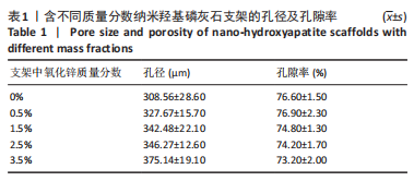
2.1 各组支架的物理性能表征 扫描电镜下可见,各组支架面具有三维贯通的孔隙,孔径大小为300-400 μm,且孔隙表面有大量微孔,见图1。5组支架的孔隙率均在70%以上,其中,0.5组的孔隙率最高,见表1。"
"
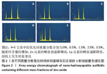
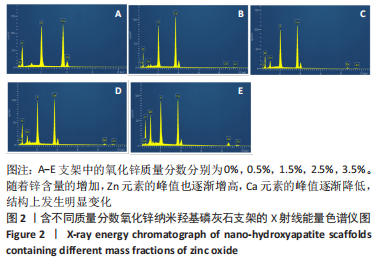
X射线能量色谱仪分析:结果显示,复合支架主要由O、Zn、Ca、P 4种元素组成,说明支架制备成功,未掺入杂质;随着锌含量的增加,Zn元素的峰值也逐渐增高,Ca元素的峰值逐渐降低,结构上发生明显变化,见图2,提示锌元素可能通过取代钙的位置掺杂到纳米羟基磷灰石中[18]。"
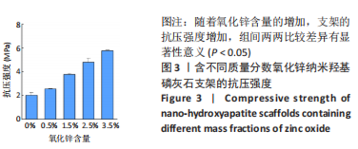
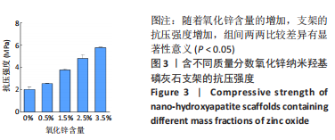
力学实验:想要修复骨缺损,复合支架的必备条件之一就是具有良好的力学性能,抗压强度是评价力学性能优劣的重要指标。纳米羟基磷灰石-氧化锌复合支架的抗压强度随着氧化锌含量的增加而增加(图3),在2.53-5.89 MPa之间,表明氧化锌可提高纳米羟基磷灰石的抗压强度,人体松质骨的抗压强度在2-11.61 MPa之间[19],表明实验制备的纳米羟基磷灰石-氧化锌复合支架满足松质骨修复材料的基本力学要求。抗压强度由大到小的顺序为:3.5%组> 2.5%组> 1.5%组> 0.5%组> 0%组,组间比较差异均有显著性意义(P < 0.05)。"
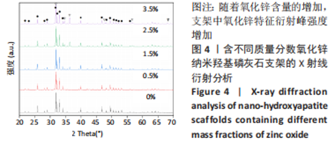
X射线衍射分析:烧结处理后的各组支架X射线衍射分析图谱显示,纳米羟基磷灰石样品位于25.928°,31.820°,40.128°,47.018°和48.738°的特征衍射峰与标准衍射图样(JCPDS Card 00-034-0010)具有可容许的匹配关系,说明尽管进行高温烧结处理后,纳米羟基磷灰石仍保持其六角形结构[20];纳米羟基磷灰石-氧化锌复合支架存在氧化锌的特征衍射峰,2θ=31.88°,35.3°,36.4°,47.5°,56.7°,68.1°,其峰值强度随着氧化锌含量的增加逐渐增加,见图4。"


2.2 各组支架对成骨细胞的生物学影响 Calcein-AM/PI活死细胞染色:MC3T3-E1成骨细胞在各组支架表面接种1 d后的活死细胞染色结果显示,成骨细胞在各组支架上生长良好,仅存在极少数死亡细胞,见图5,说明支架没有明显的细胞毒性。其中2.5%组细胞存活率高于0%组(P < 0.05),3.5%组细胞存活率低于0%组(P < 0.05),其余组间比较差异无显著性意义(P > 0.05)。"

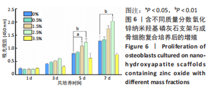
CCK-8细胞增殖实验:由图6可见,共培养的第1天,各组细胞无明显增殖;第3天,可看到0%组、1.5%组、2.5%组细胞增殖较明显;第5天,各组细胞显著增殖,0%组、1.5%组、2.5%组细胞持续增殖,进入对数生长期;第7天,各组成骨细胞的增殖表现出支架依赖性。各组复合支架的促成骨细胞活性能力依次为:2.5%组> 1.5%组> 0.5%组> 0%组> 3.5%组,与之前的锌掺入纳米羟基磷灰石可促进成骨细胞黏附、增殖和扩散的研究结论一致[21]。"

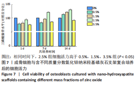
CellTiter-LumiTM发光实验:检测结果显示,随着培养时间的延长,各组成骨细胞活力均有不同程度的增强;在相同时间下(3,5,7 d),当氧化锌质量分数在0.5%-2.5%时,支架上成骨细胞的活力随氧化锌含量的增加而升高;当氧化锌质量分数为3.5%时,支架上成骨细胞的活力明显降低,见图7,说明氧化锌的掺杂有利于细胞的黏附和增殖,但超过一定含量限度会对细胞产生抑制作用。"


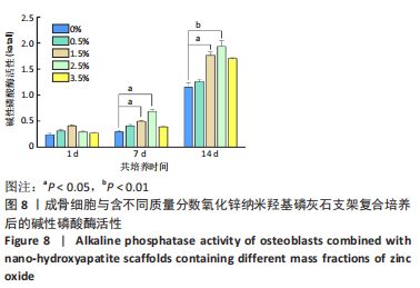
碱性磷酸酶活性检测:碱性磷酸酶是一种成骨相关因子,通过水解焦磷酸盐在促进骨矿化过程中发挥着重要作用[22]。共培养1 d后,各组细胞碱性磷酸酶活性比较差异无显著性意义(P > 0.05);共培养7,14 d后,1.5%,2.5%组细胞碱性磷酸酶活性高于0%组(P < 0.05,P < 0.01),见图8,说明氧化锌的掺杂可提高成骨细胞的碱性磷酸酶活性。这与上面的实验结果相吻合,推测该结果出现的原因是:锌为人类和动物骨骼中胶原合成和矿化所必需的[23],为碱性磷酸酶的辅基,因此锌可提高碱性磷酸酶活性,从而促进成骨。"
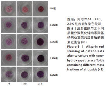
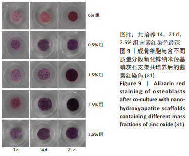
茜素红染色:在成骨细胞分化的后期,钙化结节的出现提示矿化形成,茜素红与钙离子结合使钙结节着色,从而判断骨组织矿化及成骨分化的程度。共培养7 d后,各组染色结果无明显差异,结合上述实验结果,推测7 d时成骨细胞正处于增殖期,尚未启动分化;共培养14 d后,2.5%组最有利于成骨细胞的晚期分化;共培养21 d后,茜素红染色结果与14 d时表现出的结果相似,且组间差距更明显,见图9。由于茜素红染色存在支架的干扰性,晚期可能会使实验结果观察产生偏差,因此需要钙离子定量分析验证。"
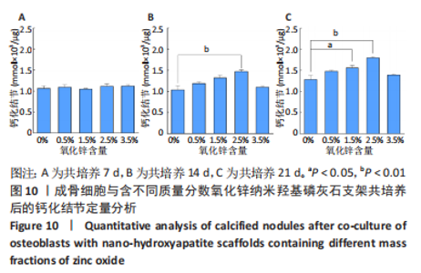
钙离子定量分析:共培养7 d后,各组钙离子含量比较差异无显著性意义(P > 0.05);共培养14 d后,2.5%组钙含量高于0%组(P < 0.05);共培养21 d后,1.5%,2.5%组钙含量高于0%组(P < 0.05,P < 0.01),见图10,说明含质量分数2.5%氧化锌的纳米羟基磷灰石支架最有利于成骨细胞的晚期分化。"


2.3 含氧化锌纳米羟基磷灰石支架的生物相容性 由体外细胞实验结果可知,含质量分数2.5%氧化锌的纳米羟基磷灰石支架具有良好的生物相容性。"

| [1] WU Y, LI Q, XU B, et al. Nano-hydroxyapatite coated TiO2 nanotubes on Ti-19Zr-10Nb-1Fe alloy promotes osteogenesis in vitro. Colloids Surf B Biointerfaces. 2021;207:112019. [2] RESENDE RFB, FERNANDES GVO, SANTOS SRA, et al. Long-term biocompatibility evaluation of 0.5% zinc containing hydroxyapatite in rabbits. J Mater Sci Mater Med. 2013;24(6):1455-1463. [3] Xu T, He X, Chen Z, et al. Effect of magnesium particle fraction on osteoinduction of hydroxyapatite sphere-based scaffolds. J Mater Chem B. 2019;7(37):5648-5660. [4] 刘洋.骨组织工程支架材料研究进展[J].临床口腔医学杂志,2019, 35(10):637-639. [5] 刘旭妍,吴兆玉,王冉旭,等.不同锌含量Zn/PLGA/HA复合支架对人牙髓干细胞粘附能力影响的体外研究[J].口腔医学研究,2018, 34(2):169-171. [6] SONG Y, WU H, GAO Y, et al. Zinc Silicate/Nano-Hydroxyapatite/Collagen Scaffolds Promote Angiogenesis and Bone Regeneration via the p38 MAPK Pathway in Activated Monocytes. ACS Appl Mater Interfaces. 2020;12(14):16058-16075. [7] WANG C, LIU J, LIU Y, et al. Study on osteogenesis of zinc-loaded carbon nanotubes/chitosan composite biomaterials in rat skull defects. J Mater Sci Mater Med. 2020;31(2):15. [8] FERNANDES MH, ALVES MM, CEBOTARENCO M, et al. Citrate zinc hydroxyapatite nanorods with enhanced cytocompatibility and osteogenesis for bone regeneration. Mater Sci Eng C Mater Biol Appl. 2020;115:111147. [9] TOZZI G, DE MORI A, OLIVEIRA A, et al. Composite Hydrogels for Bone Regeneration. Materials (Basel). 2016;9(4):267. [10] WEBSTER TJ, MASSA-SCHLUETER EA, SMITH JL, et al. Osteoblast response to hydroxyapatite doped with divalent and trivalent cations. Biomaterials. 2004;25(11):2111-2121. [11] PROMITA B, HOWA B, ABHIJIT C, et al. Animal trial on zinc doped hydroxyapatite: A case study. JAsCerS. 2014;2(1):44-51. [12] SAHA N, KESKINBORA K, SUVACI E, et al. Sintering, microstructure, mechanical, and antimicrobial properties of HAp-ZnO biocomposites. J Biomed Mater Res B Appl Biomater. 2010;95(2):430-440. [13] BANDYOPADHYAY A, WITHEY EA, MOORE J, et al. Influence of ZnO doping in calcium phosphate ceramics. Mater Sci Eng C-Bio S. 2007;27(1):14-17. [14] XING F, CHI Z, YANG R, et al. Chitin-hydroxyapatite-collagen composite scaffolds for bone regeneration. Int J Biol Macromol. 2021;184:170-180. [15] 臧洪敏,徐东潭,陈宁杰,等.深低温冻存时限对成骨细胞生物学特性影响的实验研究[J].生物医学工程学杂志,2007,24(4):894-897. [16] JONASON JH, O’KEEFE RJ. Isolation and culture of neonatal mouse calvarial osteoblasts. Methods Mol Biol. 2014;1130:295-305. [17] YIN N, ZHU L, DING L, et al. MiR-135-5p promotes osteoblast differentiation by targeting HIF1AN in MC3T3-E1 cells. Cell Mol Biol Lett. 2019;24:51. [18] MA X, ELLIS DE. Initial stages of hydration and Zn substitution/occupation on hydroxyapatite (0001) surfaces. Biomaterials. 2008;29(3):257-265. [19] EILBAGI M, EMADI R, RAEISSI K, et al. Mechanical and cytotoxicity evaluation of nanostructured hydroxyapatite-bredigite scaffolds for bone regeneration. Mater Sci Eng C Mater Biol Appl. 2016;68:603-612. [20] MALZONE A, BOTTINO L, VELOTTI A. Idrossiapatite. Nota I: Struttura chimica e fisica [Hydroxyapatite. 1. Chemical and physical structure]. Arch Stomatol (Napoli). 1989;30(2):473-479. [21] MAVROPOULOS E, HAUSEN M, COSTA AM, et al. The impact of the RGD peptide on osteoblast adhesion and spreading on zinc-substituted hydroxyapatite surface. J Mater Sci Mater Med. 2013;24(5):1271-1283. [22] WENG JJ, SU Y. Nuclear matrix-targeting of the osteogenic factor Runx2 is essential for its recognition and activation of the alkaline phosphatase gene. Biochim Biophys Acta. 2013;1830(3):2839-2852. [23] TIFFANY AS, GRAY DL, WOODS TJ, et al. The inclusion of zinc into mineralized collagen scaffolds for craniofacial bone repair applications. Acta Biomater. 2019;93:86-96. [24] YIN S, ZHANG W, ZHANG Z, et al. Recent Advances in Scaffold Design and Material for Vascularized Tissue-Engineered Bone Regeneration. Adv Healthc Mater. 2019;8(10):e1801433. [25] CHOCHOLATA P, KULDA V, BABUSKA V. Fabrication of Scaffolds for Bone-Tissue Regeneration. Materials (Basel). 2019;12(4):568. [26] BEGAM H, KUNDU B, CHANDA A, et al. MG63 osteoblast cell response on Zn doped hydroxyapatite (HAp) with various surface features. Ceramic Int. 2017;43(4):3752-3760. [27] LASUBRAMANIAN P, STROBEL LA, KNESER U, et al. Zinc— containing bioactive glasses for bone regeneration,dental and orthopedic applications. Biomed Glass. 2015;1:51-69. [28] PARK JK, KIM YJ, YEOM J, et al. The Topographic Effect of Zinc Oxide Nanoflowers on Osteoblast Growth and Osseointegration. Adv Mater. 2010;22(43):4857-4861. [29] HASHIZUME M, YAMAGUCHI M. Stimulatory effect of beta-alanyl-L-histidinato zinc on cell proliferation is dependent on protein synthesis in osteoblastic MC3T3-E1 cells. Mol Cell Biochem. 1993;122(1):59-64. [30] KISHI S, YAMAGUCHI M. Inhibitory effect of zinc compounds on osteoclast-like cell formation in mouse marrow cultures. Biochem Pharmacol. 1994;48(6):1225-1230. [31] SHUM A, LI J, MAK A, et al. Fabrication and structural characterization of porous biodegradable poly(dllacticcoglycolic acid) scaffolds with controlled range of pore sizes. Polym Degrad Stabil. 2005;87(3):487-493. [32] ZHAO J, DUAN K, ZHANG JW, et al. Preparation of highly interconnected porous hydroxyapatite scaffolds by chitin gel-casting. Mater Sci Eng C. 2011;31(3):697-701. [33] KOROLEVA MY, GORBACHEVSKI OS, YURTOV EV. Paraffin wax emulsions stabilized with polymers, surfactants, and nanoparticles. Theoret Found Cheml Eng. 2017;51(1):125-132. [34] TERRA J, JIANG M, ELLIS DE. Characterization of electronic structure and bonding in hydroxyapatite: Zn substitution for Ca. Philosophical Magazine A. 2002;82(11):2357-2377. [35] MATSUNAGA K. First-Principles Study of Substitutional Magnesium and Zinc in Hydroxyapatite and Octacalcium Phosphate. J Chem Phys. 2008;128(24):245101. [36] TANG Y, CHAPPELL HF, DOVE MT, et al. Zinc incorporation into hydroxylapatite. Biomaterials. 2009;30(15):2864-2872. [37] GUPTA D, VASHISTH P, BELLARE J. Multiscale porosity in a 3D printed gellan-gelatin composite for bone tissue engineering. Biomed Mater. 2021;16(3). doi:10.1088/1748-605X/abf1a7. [38] CHANG BS, LEE CK, HONG KS, et al. Osteoconduction at porous hydroxyapatite with various pore configurations. Biomaterials. 2000; 21(12):1291-1298. [39] KARAGEORGIOU V, KAPLAN D. Porosity of 3D biomaterial scaffolds and osteogenesis. Biomaterials. 2005;26(27):5474-5491. [40] HANNINK G, ARTS JJ. Bioresorbability, porosity and mechanical strength of bone substitutes: what is optimal for bone regeneration? Injury. 2011;42(2):S22-S25. [41] YAMAGUCHI M. Role of nutritional zinc in the prevention of osteoporosis. Mol Cell Biochem. 2010;338(1-2):241-254. [42] CHEN QZ, THOMPSON ID, BOCCACCINI AR. 45S5 Bioglass-derived glass-ceramic scaffolds for bone tissue engineering. Biomaterials. 2006; 27(11):2414-2425. [43] SAFARI GEZAZ M, MOHAMMADI AREF S, KHATAMIAN M. Investigation of structural properties of hydroxyapatite/ zinc oxide nanocomposites; an alternative candidate for replacement in recovery of bones in load-tolerating areas. Mater Chem Phys. 2019;226. [44] XUE W, DAHLQUIST K, BANERJEE A, et al. Synthesis and characterization of tricalcium phosphate with Zn and Mg based dopants. J Mater Sci Mater Med. 2008;19(7):2669-2677. [45] 罗雯静,赵静辉,孟星,等.钙磷涂层中掺入锌对成骨的影响及其潜在生物学机制[J].生物医学工程学杂志,2015,32(6):1359-1363. |
| [1] | Zhang Tingting, Liu Juan, Zhang Xu. Bioactivity of phase-transition lysozyme for surface modification of zirconia all-ceramic implant material mediating hydroxyapatite coating [J]. Chinese Journal of Tissue Engineering Research, 2023, 27(7): 1043-1049. |
| [2] | Yang Yitian, Wang Lu, Yao Wei, Zhao Bin. Application of the interaction between biological scaffolds and macrophages in bone regeneration [J]. Chinese Journal of Tissue Engineering Research, 2023, 27(7): 1071-1079. |
| [3] | Li Cheng, Zheng Guoshuang, Kuai Xiandong, Yu Weiting. Alginate scaffold in articular cartilage repair [J]. Chinese Journal of Tissue Engineering Research, 2023, 27(7): 1080-1088. |
| [4] | Tang Haotian, Liao Rongdong, Tian Jing. Application and design of piezoelectric materials for bone defect repair [J]. Chinese Journal of Tissue Engineering Research, 2023, 27(7): 1117-1125. |
| [5] | Xu Yan, Li Ping, Lai Chunhua, Zhu Peijun, Yang Shuo, Xu Shulan. Piezoelectric materials for vascularized bone regeneration [J]. Chinese Journal of Tissue Engineering Research, 2023, 27(7): 1126-1132. |
| [6] | Qin Yuxing, Ren Qiangui, Li Zilong, Quan Jiaxing, Shen Peifeng, Sun Tao, Wang Haoyu. Action mechanism and prospect of bone microvascular endothelial cells for treating femoral head necrosis [J]. Chinese Journal of Tissue Engineering Research, 2023, 27(6): 955-961. |
| [7] | Zhang Min, Zhang Xiaoming, Liu Tongbin. Application potential of naringin in bone tissue regeneration [J]. Chinese Journal of Tissue Engineering Research, 2023, 27(5): 787-792. |
| [8] | Zhou Jie, Ye Peng, Zhang Tianxi, Li Xingyu, Li Shasha, Yu Anyong, Deng Jiang. Repair of rabbit cartilage defects with double-layer bionic scaffold loaded with nerve growth factor cartilage and subchondral bone [J]. Chinese Journal of Tissue Engineering Research, 2023, 27(34): 5421-5429. |
| [9] | Teng Jianxiang, Zhu Jisheng, Yuan Daizhu, Wang Zhen, Zhou Yuhu, Tian Xiaobin. Preparation of cartilage decellularized extracellular matrix-loaded composite nanofiber scaffolds based on two-nozzle electrospinning [J]. Chinese Journal of Tissue Engineering Research, 2023, 27(34): 5448-5454. |
| [10] | Li Li, Li Xiao, Li Duchenhui, Zhang Jie, Xiao Tianjiao, Kang Jiabing, Tian Ai. Regulation of interleukin-4 on osteoclast differentiation during bone regeneration guided by bone replacement materials [J]. Chinese Journal of Tissue Engineering Research, 2023, 27(34): 5455-5461. |
| [11] | Li Xinlun, Zhu Yushu, Yang Yiling, He Siqi, Wen Nan, Mu Yandong. Construction and in vivo osteogenesis of microspheres loaded with immunomodulatory peptide/miR-26a complexes for slow release [J]. Chinese Journal of Tissue Engineering Research, 2023, 27(34): 5469-5476. |
| [12] | Zhou Yuebin, Guo Honggang. Construction of tissue-engineered bone composite scaffolds by loading rabbit-derived bone marrow mesenchymal stem cells on magnesium-based alloy scaffolds [J]. Chinese Journal of Tissue Engineering Research, 2023, 27(34): 5491-5496. |
| [13] | Zhao Kang, Lin Si, Ge Xiaotian, Du Xinrui, Han Yingchao. Biosafety evaluation of poly(lactic acid)/nano-hydroxyapatite composite microspheres [J]. Chinese Journal of Tissue Engineering Research, 2023, 27(34): 5497-5504. |
| [14] | You Yan, Chen Jiawen, Lin Binbin, Wu Jingyi, Liu Peng, Wu Buling, Sun Tianyu. Mechanism and application of glycosaminoglycan in bone tissue engineering [J]. Chinese Journal of Tissue Engineering Research, 2023, 27(34): 5538-5545. |
| [15] | Xu Zhengyi, Wan Qianbing, Chen Junyu. Natural small molecular compounds in the treatment of bone-related diseases by regulating type H blood vessels and its application in tissue engineering [J]. Chinese Journal of Tissue Engineering Research, 2023, 27(34): 5546-5553. |
| Viewed | ||||||
|
Full text |
|
|||||
|
Abstract |
|
|||||