Chinese Journal of Tissue Engineering Research ›› 2023, Vol. 27 ›› Issue (34): 5421-5429.doi: 10.12307/2023.804
Previous Articles Next Articles
Repair of rabbit cartilage defects with double-layer bionic scaffold loaded with nerve growth factor cartilage and subchondral bone
Zhou Jie1, Ye Peng1, Zhang Tianxi1, Li Xingyu1, Li Shasha1, Yu Anyong1, Deng Jiang2
- 1Emergency Department of Affiliated Hospital of Zunyi Medical University, Zunyi 563003, Guizhou Province, China; 2Department of Orthopedics, Zunyi First People’s Hospital, Zunyi 563003, Guizhou Province, China
-
Received:2022-09-17Accepted:2022-10-29Online:2023-12-08Published:2023-04-20 -
Contact:Ye Peng, MD, Associate chief physician, Emergency Department of Affiliated Hospital of Zunyi Medical University, Zunyi 563003, Guizhou Province, China -
About author:Zhou Jie, Master, Emergency Department of Affiliated Hospital of Zunyi Medical University, Zunyi 563003, Guizhou Province, China -
Supported by:National Natural Science Foundation of China (H0606) Regional Science Fund (to DJ); Guizhou Provincial Science and Technology Plan Project, No. [2021]074 (to YP); Zunyi Science and Technology Joint Fund, No. HZ(2020)247 (to YP); Doctoral Initiation Fund of Zunyi Medical University, No. (2018)02 (to YP)
CLC Number:
Cite this article
Zhou Jie, Ye Peng, Zhang Tianxi, Li Xingyu, Li Shasha, Yu Anyong, Deng Jiang. Repair of rabbit cartilage defects with double-layer bionic scaffold loaded with nerve growth factor cartilage and subchondral bone[J]. Chinese Journal of Tissue Engineering Research, 2023, 27(34): 5421-5429.
share this article
Add to citation manager EndNote|Reference Manager|ProCite|BibTeX|RefWorks

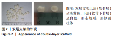
2.1 双层支架的物理性能 2.1.1 双层支架的大体观察 如图2所示,双层支架上层(软骨层)呈淡黄色,下层(软骨下骨层)呈白色,形态规则,形似圆柱体,无明显气味,质量极轻,支架长径约5 mm(软骨层约1 mm,软骨下层约4 mm),宽径4.0-5.0 mm,触之有一定弹性,弯折后不会裂开,具有良好的力学性能。"
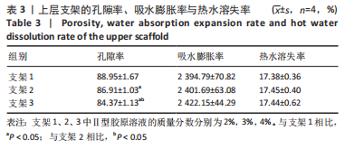
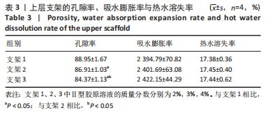
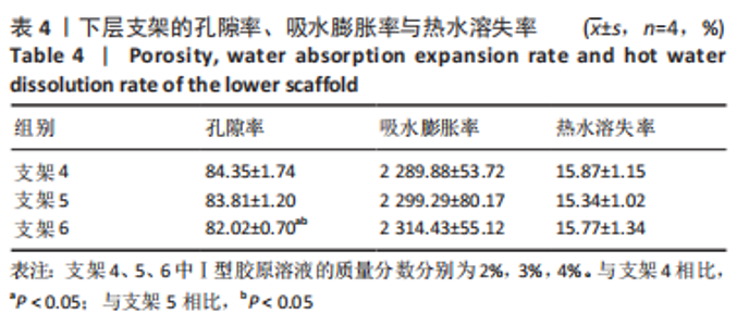
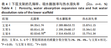
2.1.2 双层支架的孔隙率、吸水膨胀率及热水溶失率 上层支架中,3组支架的吸水膨胀率、热水溶失率比较无显著性意义(P > 0.05),孔隙率比较差异有显著性意义(P < 0.05),见表3。下层支架中,3组支架的吸水膨胀率、热水溶失率比较无显著性意义(P > 0.05),孔隙率比较差异有显著性意义(P < 0.05),见表4。"
"
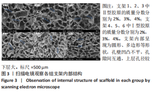
2.1.3 双层支架形态及孔径大小 如图3所示,扫描电镜显示支架内部呈现为圆形、多边形等形状,孔壁凹凸不平,孔隙间互通,上层支架1、2、3的平均孔径分别为(271.31±23.29),(268.31±22.36),(269.14±10.24) μm,各组间对比差异无显著性意义(F=0.063,P=0.939,n=10);下层支架4、5、6的平均孔径分别为(190.24±13.73),(188.45±12.73),(192.57±8.35) μm,各组间对比差异无显著性意义(F=0.274,P=0.761,n=10)。上层支架整体平均孔径为(269.08±20.49) μm,下层支架整体平均孔径为(190.42±11.96) μm,上层孔径较下层更大(P < 0.05)。"

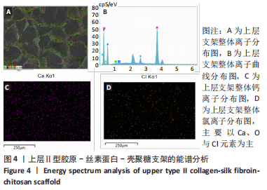
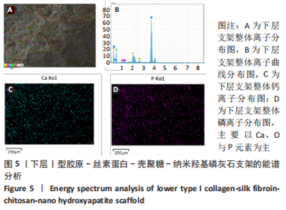
2.1.4 能谱分析及离子分布检测 如图4,5所示,支架上层(软骨层)主要以Ca、O与Cl元素为主,下层(软骨下层)主要以Ca、O与P元素为主,结果显示支架表面有羟基磷灰石的沉积,能为后期骨组织矿化形成提供原材料,具有有良好的生物活性。"
"

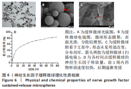
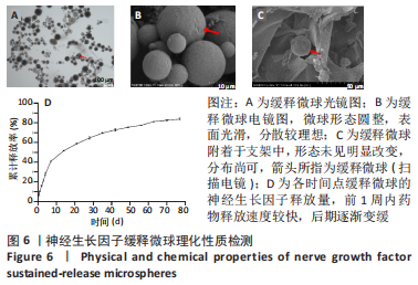
2.1.5 支架筛选结果 由以上结果,最后选择支架1与支架4进行黏合,进行后续实验。 2.2 缓释微球表征结果 缓释微球形态圆整,表面光滑,分散较理想,平均粒径为(25.87±6.54) μm,见图6A、B,其载药量为0.516 μg/mg,包封率为40.5%;微球与支架结合后未见明显微球丢失及突释,分布尚可,见图6C。神经生长因子缓释微球体外释放检测显示,在前1周内药物释放速度较快,第1天神经生长因子微球释放量为(10.67±0.42)%,第7天为(40.87±0.55)%,后期逐渐变缓,实验结果达到了控制药物缓慢释放,提高药物生物利用率的目的,见图6D。"

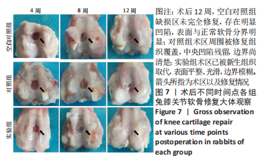
2.3 双层支架兔软骨缺损修复效果 2.3.1 实验动物数量分析 30只新西兰大白兔均存活,术后第1-3天术区及关节肿胀,活动以及饮食欠佳;术后1周术区及关节处肿胀明显消退,活动较前明显好转。30只兔全部进入结果分析。 2.3.2 软骨大体学观察及ICRS软骨修复评分 关节软骨大体学观察(图7):术后标本提取时发现,空白对照组关节少许粘连,滑囊呈现轻度充血水肿,其余两组动物关节及滑囊正常,没有明显滑膜炎症、骨赘形成。空白对照组术后4,8,12周缺损区未完全修复,存在明显凹陷,表面与正常软骨分界明显。对照组术后4周见术区凹陷明显;术后8周时术区被部分的组织覆盖,凹陷区域变平;术后12周时术区周围被修复组织覆盖,中央凹陷残留,边界尚清楚。实验组术后4周尚可见缺损区凹陷轮廓;术后8周时术区被修复组织填充,凹陷区域明显变平;术后12周时术区已被新生组织取代,表面平整、光滑,边界模糊。"
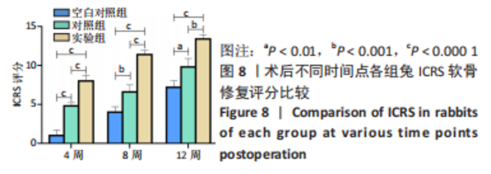
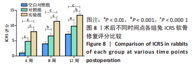
ICRS软骨修复评分:3组ICRS软骨修复评分随术后时间逐渐递增,各组内、组间对比差异均有显著性意义(P < 0.05)。实验组各时间段评分高于对照组、空白对照组(P < 0.05),对照组各时间段评分高于空白对照组(P < 0.05),见图8。"
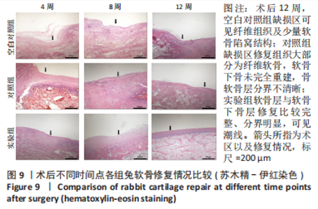
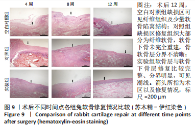
2.3.3 组织学观察及Wakitani病理评分 苏木精-伊红染色(图9):空白对照组术后4周缺损区未见骨组织再生;术后8周缺损区可见部分纤维组织及骨重建;术后12周缺损区可见纤维组织及少量软骨陷窝结构。对照组术后4周缺损区以纤维组织为主;术后8周缺损区软骨层纤维组织增生,软骨下骨层出现部分骨小梁结构,分界不清;术后12周缺损区修复组织大部分为纤维软骨,软骨下骨未完全重建,骨软骨层分界不清晰。实验组术后4周缺损区可见纤维软骨修复;术后8周缺损区类透明软骨增生明显,较多骨小梁结构可见,未出现潮线;术后12周软骨层与软骨下骨层修复比较完整、分界明显,可见潮线。"
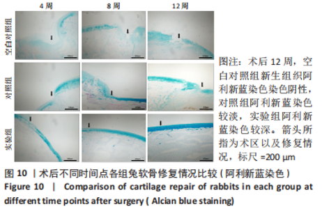
阿利新蓝染色(图10):空白对照组术后4-12周时缺损区仍可见明显软骨缺失,新生组织阿利新蓝染色染色阴性。对照组术后4-12周缺损区逐渐被新生组织填充,表面变光滑,与周围软骨连续,阿利新蓝染色较淡。实验组术后4-12周缺损区被新生组织填充,表面光滑,阿利新蓝染色由淡染逐渐变为深染色,骨软骨层可见明显分界。"


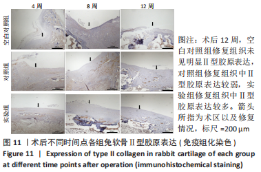
Ⅱ型胶原免疫组化染色(图11):空白对照组修复组织术后12周仍未见明显Ⅱ型胶原表达。对照组到术后8周时修复组织中可见Ⅱ型胶原少量表达,12周时修复组织中Ⅱ型胶原表达较弱。实验组术后4,8周修复软骨Ⅱ型胶原均有表达,12周时修复组织中Ⅱ型胶原表达更多,骨软骨分界明显,可见潮线。"

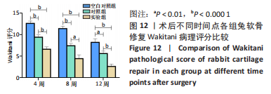
Wakitani病理评分:3组Wakitani病理评分随术后时间的延长逐渐降低,各组内、组间对比差异均有显著性意义(P < 0.05)。实验组各时间点评分明显低于对照组、空白对照组(P < 0.05),对照组各时间点评分低于空白对照组(P < 0.05),见图12。"

2.3.4 双层支架的组织相容性 由动物实验可知,双层支架具有良好的组织相容性。 "

| [1] BEATA Ż, ALEKSANDRA SR, URSZULA L, et al. Structure and Pathologies of Articular Cartilage. In Vivo. 2021;35:1355-1363. [2] ABHIJIT MB, JAMES BR. Articular cartilage: structure, injuries and review of management. Br Med Bull. 2008;87:77-95. [3] JOSÉ B, JOSÉ AA, ENRIQUE G, et al. Articular cartilage: structure and regeneration. Tissue Eng Part B Rev. 2010;16:617-627. [4] MATTHEW JK, JOHN WB, JUSTIN MP, et al. Microfracture Versus Autologous Chondrocyte Implantation for Articular Cartilage Lesions in the Knee: A Systematic Review of 5-Year Outcomes. Am J Sports Med. 2018;46:995-999. [5] REISSIS D, TANG QO, COOPER NC, et al. Current clinical evidence for the use of mesenchymal stem cells in articular cartilage repair. Expert Opin Biol Ther. 2016; 16(4):535-557. [6] XIN L, ZHANG C, XU WX, et al. Current advances on surgical treatment for knee articular cartilage injuries. Zhongguo Gu Shang. 2018;31:281-285. [7] HÖNLE W, JEZUSSEK D, FABIJANI R, et al. Surgical treatment of knee osteoarthritis. MMW Fortschr Med. 2007;149:33-34,36. [8] CHASE SD, JORGE C, CRUZ RS, et al. Fresh Osteochondral Allograft Transplantation for Treatment of Articular Cartilage Defects of the Knee. Arthrosc Tech. 2016;5: e157-161. [9] EREN Y, ALI E, EMRE Y, et al. The Effect of Intraarticular Insulin on Chondral Defect Repair. Cartilage. 2021;13:684S-691S. [10] RIMTAUTAS G, AGNE G, ARNOLDAS P, et al. Ten-year follow-up of a prospective, randomized clinical study of mosaic osteochondral autologous transplantation versus microfracture for the treatment of osteochondral defects in the knee joint of athletes. Am J Sports Med. 2012;40:2499-2508. [11] SAHAR J, SADRABADI AMIN E, FATEMEH Z, et al. New Insights into Cartilage Tissue Engineering: Improvement of Tissue-Scaffold Integration to Enhance Cartilage Regeneration. Biomed Res Int. 2022;2022:7638245. [12] 杨东翔,元香南,孔战,等.组织工程软骨的研究现状与进展[J].山东医药, 2011,51(40):113-114. [13] HOLZAPFEL BM, RUDERT M, HUTMACHER DW. Scaffold-based Bone Tissue Engineering. Orthopade. 2017;46:701-710. [14] ABIY W, ELENI KT, CAGRI A, et al. Current state of fabrication technologies and materials for bone tissue engineering. Acta Biomater. 2018;80:1-30. [15] 张佼佼,王冬青,王志强,等.组织工程软骨支架材料的应用选择[J].中国组织工程研究与临床康复,2011,15(29):5467-5470. [16] XIAO HL, HUANG WL, XIONG K, et al. Osteochondral repair using scaffolds with gradient pore sizes constructed with silk fibroin, chitosan, and nano-hydroxyapatite. Int J Nanomedicine. 2019;14:2011-2027. [17] LU ZH, LEI DQ, JIANG TM, et al. Nerve growth factor from Chinese cobra venom stimulates chondrogenic differentiation of mesenchymal stem cells. Cell Death Dis. 2017;8:e2801. [18] ZHAN XT, CAI PA, LEI DQ, et al. Comparative profiling of chondrogenic differentiation of mesenchymal stem cells (MSCs) driven by two different growth factors. Cell Biochem Funct. 2019;37:359-367. [19] JI L, SONG ZW, ZENG FH, et al. Research progress on controlled release of various growth factors in bone regeneration. Zhongguo Xiu Fu Chong Jian Wai Ke Za Zhi. 2019;33:750-755. [20] CHEN ZJ, CHENG Q, YU XF, et al. Research progress of growth factor sustained -release microspheres in fat transplantation. Zhongguo Xiu Fu Chong Jian Wai Ke Za Zhi. 2017;31:1402-1406. [21] 王富友.组织工程骨软骨仿生设计与初步构建[D].重庆:第三军医大学,2008. [22] LEVINGSTONE TJ, THOMPSON E, MATSIKO A, et al. Multi-layered collagenbased scaffolds for osteochondral defect repair in rabbits. Acta Biomater. 2016;32:149-160. [23] LEVINGSTONE TJ, MATSIKO A, DICKSON GR, et al. A biomimetic multilayered collagen-based scaffold for osteochondral repair. Acta Biomater. 2014;10:1996-2004. [24] FU LW, YANG Z, GAO CJ, et al. Advances and prospects in biomimetic multilayered scaffolds for articular cartilage regeneration. Regen Biomater. 2020;7:527-542. [25] 康嘉宇,吕建伟,赵志虎,等.骨软骨组织工程分层支架的研究进展[J].中华骨科杂志,2019,39(22):1413-1420. [26] PEREIRA DR, RUI L REIS RL, OLIVEIRA JM. Layered Scaffolds for Osteochondral Tissue Engineering. Adv Exp Med Biol. 2018;1058:193-218. [27] IVANA G, GORDAN VN.Challenges in engineering osteochondral tissue grafts with hierarchical structures. Expert Opin Biol Ther. 2015;15:1583-1599. [28] 朱浩方,毛宏理,顾忠伟.医用组织粘合剂的研究进展[J].中国材料进展,2020, 39(Z1):535-550+557-558. [29] KAZUNORI S, YU M, CHRISTOPHER DM, et al. Osteochondral tissue engineering with biphasic scaffold: current strategies and techniques. Tissue Eng Part B Rev. 2014; 20:468-476. [30] PROMITA B, BANANI K, DEBOKI N, et al. Silk scaffolds in bone tissue engineering: An overview. Acta Biomater. 2017;63:1-17. [31] WALTER B, WEERASAK S, ARAMWIT P, et al. Natural Origin Materials for Osteochondral Tissue Engineering. Adv Exp Med Biol. 2018;1058:3-30. [32] MIRIAM F, GORDIAN B, MANSOOR C, et al. Natural Polymeric Scaffolds in Bone Regeneration. Front Bioeng Biotechnol. 2020;8:474. [33] JOANNE R, JOHN R, KEVIN MS, et al. Biomaterials and scaffold design: key to tissue-engineering cartilage. Biotechnol Appl Biochem. 2007;46:73-84. [34] DEL BAKHSHAYESH AR, ASADI N, ALIHEMMATI A, et al. An overview of advanced biocompatible and biomimetic materials for creation of replacement structures in the musculoskeletal systems: focusing on cartilage tissue engineering. J Biol Eng. 2019;13:85. [35] NAZANIN A, MARZIYEH F, NOROOZI PN, et al. Bioactive polymeric scaffolds for osteogenic repair and bone regenerative medicine. Med Res Rev. 2020;40:1833-1870. [36] MANO JF, REIS RL. Osteochondral defects: present situation and tissue engineering approaches. J Tissue Eng Regen Med. 2007;1:261-273. [37] HUTMACHER DW. Scaffolds in tissue engineering bone and cartilage. Biomaterials. 2000;21:2529-2543. [38] SMITH GD, KNUTSEN G, RICHARDSON JB. A clinical review of cartilage repair techniques. J Bone Joint Surg Br. 2005;87:445-449. [39] TANYA JL, AMOS M, GLENN RD, et al. A biomimetic multi-layered collagen-based scaffold for osteochondral repair. Acta Biomater. 2014;10:1996-2004. [40] MASTROGIACOMO M, MURAGLIA A, KOMLEV V, et al. Tissue engineering of bone: search for a better scaffold. Orthod Craniofac Res. 2005;8:277-284. [41] 高泽,石志良,李锋,等.材料和孔隙率对可降解支架内骨形成的影响[J].医用生物力学,2021,36(4):582-588. [42] LAMINE C, JÉRÔME C, MARION D, et al. Autologous osteochondral mosaicplasty in osteochondritis dissecans of the patella in adolescents. Int Orthop. 2017;41:197-202. [43] AARON JK, HEATHER WH, SCOTT AR, et al. Activity levels are higher after osteochondral autograft transfer mosaicplasty than after microfracture for articular cartilage defects of the knee: a retrospective comparative study. J Bone Joint Surg Am. 2012;94:971-978. [44] MARCUS M, ANDREA B, SYLVIE M, et al. Nasal chondrocyte-based engineered autologous cartilage tissue for repair of articular cartilage defects: an observational first-in-human trial. Lancet. 2016;388:1985-1994. [45] MAURILIO M, ELIZAVETA K, MARCO D, et al. Arthroscopic autologous osteochondral grafting for cartilage defects of the knee: prospective study results at a minimum 7-year follow-up. Am J Sports Med. 2007;35:2014-2021. [46] WIDYASRI P, YOSHIHITO N, SILVIA G, et al. Bone ingrowth of various porous titanium scaffolds produced by a moldless and space holder technique: an in vivo study in rabbits. Biomed Mater. 2016;11:015012. [47] ORTH M, FRITZ T, STUTZ J, et al. Local Application of Mineral-Coated Microparticles Loaded With VEGF and BMP-2 Induces the Healing of Murine Atrophic Non-Unions. Front Bioeng Biotechnol. 2021;9:809397. [48] MIKAËL MM, PRISCILLA SB, KENTA M, et al. Extracellular matrix-inspired growth factor delivery systems for bone regeneration. Adv Drug Deliv Rev. 2015;94:41-52. [49] VALENTINA D, ELISA L, GABRIELA C, et al. Growth factors in bone repair. Chir Organi Mov. 2008;92:161-168. [50] 刘华蔚.壳聚糖导管复合NGF缓释微球修复兔面神经缺损的实验研究[D].北京:中国人民解放军军医进修学院,2011. [51] ZHANG JX, ZHU KJ, CHEN D. Preparation of bovine serum albumin loaded poly (D, L-lactic -co-glycolic acid) microspheres by a modified phase separation technique. J Microencapsul. 2005;22:117-126. [52] KEN Y, KATHY T, SHALLEY AR, et al. Osteochondral repair using an acellular dermal matrix-pilot in vivo study in a rabbit osteochondral defect model. J Orthop Res. 2018;36:1919-1928. [53] TROY DB, ADETOLA BA, NADR MJ, et al. Articular Cartilage Repair with Mesenchymal Stem Cells After Chondrogenic Priming: A Pilot Study. Tissue Eng Part A. 2018;24: 761-774. [54] HUANG Y, FAN HQ, GONG XY, et al. Scaffold With Natural Calcified Cartilage Zone for Osteochondral Defect Repair in Minipigs .Am J Sports Med. 2021;49:1883-1891. [55] IRINA AD M, RAFAEL AVB, HAROLD B, et al. Fixation of Hydrogel Constructs for Cartilage Repair in the Equine Model: A Challenging Issue. Tissue Eng Part C Methods. 2017;23:804-814. [56] FREED LE, GRANDE DA, LINGBIN Z, et al. Joint resurfacing using allograft chondrocytes and synthetic biodegradable polymer scaffolds. J Biomed Mater Res. 1994;28:891-899. [57] ISABEL RD, CARLOS AV, PEDRO PC. Large Animal Models for Osteochondral Regeneration. Adv Exp Med Biol. 2018;1059:441-501. |
| [1] | Sun Kexin, Zeng Jinshi, Li Jia, Jiang Haiyue, Liu Xia. Mechanical stimulation enhances matrix formation of three-dimensional bioprinted cartilage constructs [J]. Chinese Journal of Tissue Engineering Research, 2023, 27(在线): 1-7. |
| [2] | Xu Xingxing, Wen Chaoju, Meng Maohua, Wang Qinying, Chen Jingqiao, Dong Qiang. Carbon nanomaterials in oral implant [J]. Chinese Journal of Tissue Engineering Research, 2023, 27(7): 1062-1070. |
| [3] | Yang Yitian, Wang Lu, Yao Wei, Zhao Bin. Application of the interaction between biological scaffolds and macrophages in bone regeneration [J]. Chinese Journal of Tissue Engineering Research, 2023, 27(7): 1071-1079. |
| [4] | Li Cheng, Zheng Guoshuang, Kuai Xiandong, Yu Weiting. Alginate scaffold in articular cartilage repair [J]. Chinese Journal of Tissue Engineering Research, 2023, 27(7): 1080-1088. |
| [5] | Shi Yehong, Wang Cheng, Chen Shijiu. Early thrombosis and prevention of small-diameter blood vessel prosthesis [J]. Chinese Journal of Tissue Engineering Research, 2023, 27(7): 1110-1116. |
| [6] | Tang Haotian, Liao Rongdong, Tian Jing. Application and design of piezoelectric materials for bone defect repair [J]. Chinese Journal of Tissue Engineering Research, 2023, 27(7): 1117-1125. |
| [7] | Xu Yan, Li Ping, Lai Chunhua, Zhu Peijun, Yang Shuo, Xu Shulan. Piezoelectric materials for vascularized bone regeneration [J]. Chinese Journal of Tissue Engineering Research, 2023, 27(7): 1126-1132. |
| [8] | Li Xinyue, Li Xiheng, Mao Tianjiao, Tang Liang, Li Jiang. Three-dimensional culture affects morphology, activity and osteogenic differentiation of human periodontal ligament stem cells [J]. Chinese Journal of Tissue Engineering Research, 2023, 27(6): 846-852. |
| [9] | Li Xiaoyin, Yang Xiaoqing, Chen Shulian, Li Zhengchao, Wang Ziqi, Song Zhen, Zhu Daren, Chen Xuyi. Collagen/silk fibroin scaffold combined with neural stem cells in the treatment of traumatic spinal cord injury [J]. Chinese Journal of Tissue Engineering Research, 2023, 27(6): 890-896. |
| [10] | Yuan Bo, Xie Lide, Fu Xiumei. Schwann cell-derived exosomes promote the repair and regeneration of injured peripheral nerves [J]. Chinese Journal of Tissue Engineering Research, 2023, 27(6): 935-940. |
| [11] | Qin Yuxing, Ren Qiangui, Li Zilong, Quan Jiaxing, Shen Peifeng, Sun Tao, Wang Haoyu. Action mechanism and prospect of bone microvascular endothelial cells for treating femoral head necrosis [J]. Chinese Journal of Tissue Engineering Research, 2023, 27(6): 955-961. |
| [12] | Xiong Bohan, Yu Yang, Lu Xiaojun, Wang Xu, Yang Tengyun, Zhang Yaozhang, Liao Xinyu, Zhou Xiaoxiang, He Lu, Li Yanlin. Research progress in promoting tendon to bone healing during anterior cruciate ligament reconstruction [J]. Chinese Journal of Tissue Engineering Research, 2023, 27(5): 779-786. |
| [13] | Zhang Min, Zhang Xiaoming, Liu Tongbin. Application potential of naringin in bone tissue regeneration [J]. Chinese Journal of Tissue Engineering Research, 2023, 27(5): 787-792. |
| [14] | Liu Zixuan, Li Yan, Ji Lin, Xia Delin. Biological properties of nano-hydroxyapatite-zinc oxide composite scaffolds and their effects on the behavior of MC3T3-E1 osteoblasts [J]. Chinese Journal of Tissue Engineering Research, 2023, 27(34): 5441-5447. |
| [15] | Teng Jianxiang, Zhu Jisheng, Yuan Daizhu, Wang Zhen, Zhou Yuhu, Tian Xiaobin. Preparation of cartilage decellularized extracellular matrix-loaded composite nanofiber scaffolds based on two-nozzle electrospinning [J]. Chinese Journal of Tissue Engineering Research, 2023, 27(34): 5448-5454. |
| Viewed | ||||||
|
Full text |
|
|||||
|
Abstract |
|
|||||