Chinese Journal of Tissue Engineering Research ›› 2023, Vol. 27 ›› Issue (34): 5491-5496.doi: 10.12307/2023.726
Previous Articles Next Articles
Construction of tissue-engineered bone composite scaffolds by loading rabbit-derived bone marrow mesenchymal stem cells on magnesium-based alloy scaffolds
Zhou Yuebin1, Guo Honggang2
- 1Department of Orthopedics, Tianjin Hospital, Tianjin 300200, China; 2Department of Orthopedics, General Hospital, Tianjin Medical University, Tianjin 300052, China
-
Received:2022-09-14Accepted:2022-11-16Online:2023-12-08Published:2023-04-22 -
Contact:Guo Honggang, MD, Chief physician, Department of Orthopedics, General Hospital, Tianjin Medical University, Tianjin 300052, China -
About author:Zhou Yuebin, Master, Physician, Department of Orthopedics, Tianjin Hospital, Tianjin 300200, China
CLC Number:
Cite this article
Zhou Yuebin, Guo Honggang. Construction of tissue-engineered bone composite scaffolds by loading rabbit-derived bone marrow mesenchymal stem cells on magnesium-based alloy scaffolds[J]. Chinese Journal of Tissue Engineering Research, 2023, 27(34): 5491-5496.
share this article
Add to citation manager EndNote|Reference Manager|ProCite|BibTeX|RefWorks

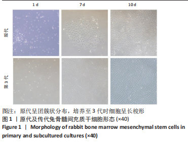
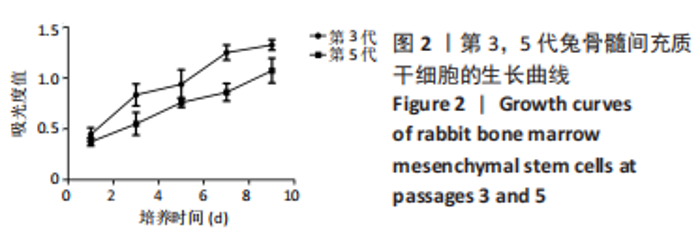
2.1 骨髓间充质干细胞生长趋势及流式细胞鉴定结果 原代及传代培养细胞形态学见图1。第3代骨髓间充质干细胞生长曲线呈典型的倒S形。传代1-3 d为生长滞缓期,第3天起细胞大量增殖,第7天达到增长峰值,此后增殖缓慢进入平台期。第5代细胞增殖能力比第3代差,生长曲线同第3代骨髓间充质干细胞,见图2。"
"

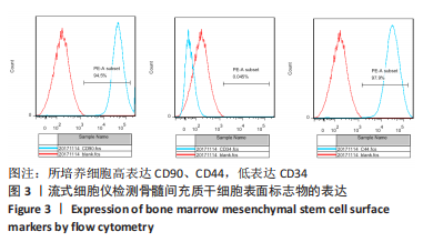
流式细胞测定显示CD34抗体阳性细胞占细胞总数的0.045%,而CD90抗体阳性细胞占细胞总数的94.5%,CD44抗体阳性细胞占细胞总数的97.9%,见图3。"
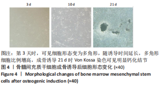
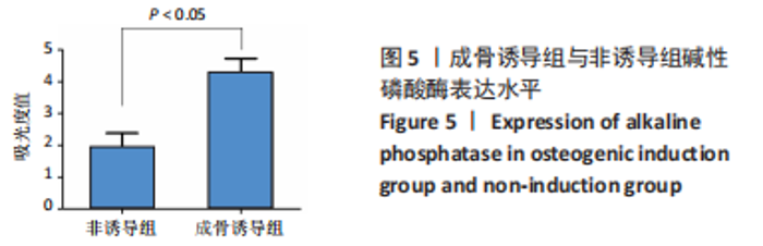
2.2 骨髓间充质干细胞成骨诱导能力 经成骨诱导的兔骨髓间充质干细胞形态发生明显变化,逐渐由长梭形变为三角形、多角形等成骨细胞样形态。倒置相差显微镜下观察:成骨诱导后第3天,部分细胞形态由长梭形逐渐变为多角形,体积增大,随诱导时间延长,多角形细胞比例增高。经成骨诱导后,Von Kossa染色可见钙化结节,见图4。成骨诱导组细胞碱性磷酸酶吸光度值为4.277±0.402,非诱导细胞组细胞碱性磷酸酶吸光度值为1.900±0.428,差异有显著性意义(P < 0.05),见图5。"

"


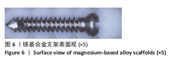
2.3 两种支架大体形貌及理化性征 2.3.1 镁基合金支架大体形貌及理化性征 镁基合金支架成分为镁、锌、钙按(96.8∶3∶0.2)分子质量分数比混合而成,统一为高8 mm,直径1.8 mm,具体形貌为具备微槽的棒状金属物,微槽间距排列规整,结构致密,见图6。镁基合金支架力学性能测试以弹性模量、抗拉强度以及延伸率为检测指标,弹性模量为45 GPa,接近骨弹性模量值,抗拉强度为336 MPa,延伸率为17.85%。"

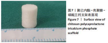
2.3.2 聚已内酯-壳聚糖-磷酸三钙支架大体形貌及理化性征 聚已内酯-壳聚糖-磷酸三钙支架为课题组前期实验所制备[14-15],其中聚已内酯与磷酸三钙质量比为2∶4,直径为10 mm,高为12 mm,为表面粗糙的圆柱状物,其弹性模量为8.0 MPa,见图7。"
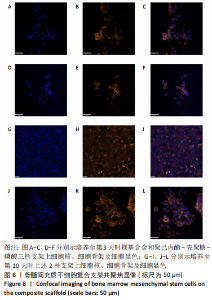
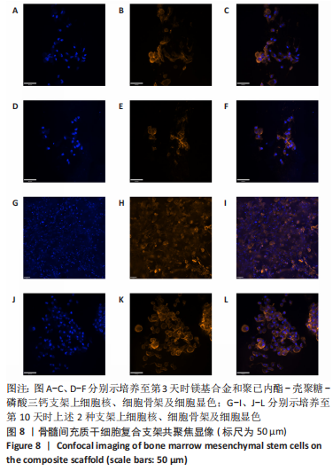
2.4 骨髓间充质干细胞与镁基合金支架共培养后激光共聚焦显微镜观察结果 骨髓间充质干细胞与镁基合金支架共培养第3天时,细胞数量与聚已内酯-壳聚糖-磷酸三钙支架相差无几,细胞黏附可,排列不规整,细胞骨架清晰,而在培养至第10天时,镁基合金支架上细胞数量明显高于聚已内酯-壳聚糖-磷酸三钙支架,镁基合金支架上细胞呈三维性生长,细胞骨架清晰,细胞核深染,排列较第3天时规整,见图8。"
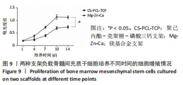
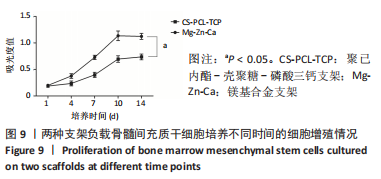
2.5 骨髓间充质干细胞与支架共培养后细胞增殖活性 在培养第1,4,7,10,14天MTT法检测细胞在490 nm处吸光度值,结果显示镁基合金支架与聚已内酯-壳聚糖-磷酸三钙支架在培养起始细胞增殖活性相差无几;随培养时间延长,镁基合金组较聚已内酯-壳聚糖-磷酸三钙组细胞增殖活性高(P < 0.05),培养至第10天左右,细胞增殖达高峰,见图9。"
| [1] LANGER R, VACANTI J. Advances in tissue engineering. J Pediatr Surg. 2016;51(1):8-12. [2] FU X, LIU G, HALIM A, et al. Mesenchymal Stem Cell Migration and Tissue Repair. Cells. 2019;8(8):784. [3] SHI Y, HU Y, LV C, et al. Effects of Reactive Oxygen Species on Differentiation of Bone Marrow Mesenchymal Stem Cells. Ann Transplant. 2016;21:695-700. [4] YANG F, LI WB, QU YW, et al. Bone marrow mesenchymal stem cells induce M2 microglia polarization through PDGF-AA/MANF signaling. World J Stem Cells. 2020;12(7):633-658. [5] YU Y, JIN G, XUE Y, et al. Multifunctions of dual Zn/Mg ion co-implanted titanium on osteogenesis, angiogenesis and bacteria inhibition for dental implants. Acta Biomater. 2017;49:590-603. [6] NASR AZADANI M, ZAHEDI A, BOWOTO OK, et al. A review of current challenges and prospects of magnesium and its alloy for bone implant applications. Prog Biomater. 2022;11(1):1-26. [7] WANG J, TANG J, ZHANG P, et al. Surface modification of magnesium alloys developed for bioabsorbable orthopedic implants: a general review. J Biomed Mater Res B Appl Biomater. 2012;100(6):1691-1701. [8] MOSTAED E, SIKORA-JASINSKA M, DRELICH JW, et al. Zinc-based alloys for degradable vascular stent applications. Acta Biomater. 2018;71:1-23. [9] LIU Y, RATH B, TINGART M, et al. Role of implants surface modification in osseointegration: A systematic review. J Biomed Mater Res A. 2020; 108(3):470-484. [10] CHA PR, HAN HS, YANG GF, et al. Biodegradability engineering of biodegradable Mg alloys: tailoring the electrochemical properties and microstructure of constituent phases. Sci Rep. 2013;3:2367. [11] KIM HK, HAN HS, LEE KS, et al. Comprehensive study on the roles of released ions from biodegradable Mg-5 wt% Ca-1 wt% Zn alloy in bone regeneration. J Tissue Eng Regen Med. 2017;11(10):2710-2724. [12] ZHANG C, LIN J, NGUYEN NT, et al. Antimicrobial Bioresorbable Mg-Zn-Ca Alloy for Bone Repair in a Comparison Study with Mg-Zn-Sr Alloy and Pure Mg. ACS Biomater Sci Eng. 2020;6(1):517-538. [13] FARTO-VAAMONDE X, AURIEMMA G, AQUINO RP, et al. Post-manufacture loading of filaments and 3D printed PLA scaffolds with prednisolone and dexamethasone for tissue regeneration applications. Eur J Pharm Biopharm. 2019;141:100-110. [14] GUO H, LI F, ZHOU YB. The Development of a Biomimetic Nanoscaled Spinal Cage Based on Three-Dimensional Reconstruction Imaging. Biomaterials and Tissue Engineering. 2016;7(6):526-530. [15] GOSSET A, BARREIRO-VILLAVERDE D, BECERRA PERMUY JC, et al. Experimental and Numerical Investigation of the Extrusion and Deposition Process of a Poly(lactic Acid) Strand with Fused Deposition Modeling. Polymers (Basel). 2020;12(12):2885. [16] CHEN Q, SHOU P, ZHENG C, et al. Fate decision of mesenchymal stem cells: adipocytes or osteoblasts? Cell Death Differ. 2016;23(7): 1128-1139. [17] LIU Z, CHANG H, HOU Y, et al. Lentivirus‑mediated microRNA‑26a overexpression in bone mesenchymal stem cells facilitates bone regeneration in bone defects of calvaria in mice. Mol Med Rep. 2018; 18(6):5317-5326. [18] KHAYAT S, FANAEI H, GHANBARZEHI A. Minerals in Pregnancy and Lactation: A Review Article. J Clin Diagn Res. 2017;11(9):QE01-QE05. [19] XUE J, SINGH S, ZHOU Y, et al. A biodegradable 3D woven magnesium-based scaffold for orthopedic implants. Biofabrication. 2022;14(3). doi: 10.1088/1758-5090/ac73b8. [20] ZHANG N, WANG W, ZHANG X, et al. The effect of different coatings on bone response and degradation behavior of porous magnesium-strontium devices in segmental defect regeneration. Bioact Mater. 2020;6(6):1765-1776. [21] QI T, WENG J, YU F, et al. Insights into the Role of Magnesium Ions in Affecting Osteogenic Differentiation of Mesenchymal Stem Cells. Biol Trace Elem Res. 2021;199(2):559-567. [22] ABED E, MOREAU R. Importance of melastatin-like transient receptor potential 7 and magnesium in the stimulation of osteoblast proliferation and migration by platelet-derived growth factor. Am J Physiol Cell Physiol. 2009;297(2):C360-C368. [23] CAI S, LEI T, LI N, et al. Effects of Zn on microstructure, mechanical properties and corrosion behavior of Mg–Zn alloys. Mater Sci Eng C Mater Biol Appl. 2012;32(8):2570-2577. [24] HE G, WU Y, ZHANG Y, et al. Addition of Zn to the ternary Mg-Ca-Sr alloys significantly improves their antibacterial property. J Mater Chem B. 2015;3(32):6676-6689. [25] SEONG JW, KIM WJ. Development of biodegradable Mg-Ca alloy sheets with enhanced strength and corrosion properties through the refinement and uniform dispersion of the Mg₂Ca phase by high-ratio differential speed rolling. Acta Biomater. 2015;11:531-542. [26] YOSHIZAWA S, BROWN A, BARCHOWSKY A, et al. Magnesium ion stimulation of bone marrow stromal cells enhances osteogenic activity, simulating the effect of magnesium alloy degradation. Acta Biomater. 2014;10(6):2834-2842. [27] WU L, FEYERABEND F, SCHILLING AF, et al. Effects of extracellular magnesium extract on the proliferation and differentiation of human osteoblasts and osteoclasts in coculture. Acta Biomater. 2015;27: 294-304. [28] ZHANG X, CHEN Q, MAO X. Magnesium Enhances Osteogenesis of BMSCs by Tuning Osteoimmunomodulation. Biomed Res Int. 2019; 2019:7908205. [29] LIU J, ZENG H, XIAO P, et al. Sustained Release of Magnesium Ions Mediated by a Dynamic Mechanical Hydrogel to Enhance BMSC Proliferation and Differentiation. ACS Omega. 2020;5(38):24477-24486. [30] WANG Z, LIU Q, LIU C, et al. Mg2+ in β-TCP/Mg-Zn composite enhances the differentiation of human bone marrow stromal cells into osteoblasts through MAPK-regulated Runx2/Osx. J Cell Physiol. 2020;235(6):5182-5191. |
| [1] | Dang Yi, Du Chengyan, Yao Honglin, Yuan Nenghua, Cao Jin, Xiong Shan, Zhang Dingmei, Wang Xin. Hormonal osteonecrosis and oxidative stress [J]. Chinese Journal of Tissue Engineering Research, 2023, 27(9): 1469-1476. |
| [2] | Yang Yitian, Wang Lu, Yao Wei, Zhao Bin. Application of the interaction between biological scaffolds and macrophages in bone regeneration [J]. Chinese Journal of Tissue Engineering Research, 2023, 27(7): 1071-1079. |
| [3] | Li Cheng, Zheng Guoshuang, Kuai Xiandong, Yu Weiting. Alginate scaffold in articular cartilage repair [J]. Chinese Journal of Tissue Engineering Research, 2023, 27(7): 1080-1088. |
| [4] | Tang Haotian, Liao Rongdong, Tian Jing. Application and design of piezoelectric materials for bone defect repair [J]. Chinese Journal of Tissue Engineering Research, 2023, 27(7): 1117-1125. |
| [5] | Liu Wentao, Feng Xingchao, Yang Yi, Bai Shengbin. Effect of M2 macrophage-derived exosomes on osteogenic differentiation of bone marrow mesenchymal stem cells [J]. Chinese Journal of Tissue Engineering Research, 2023, 27(6): 840-845. |
| [6] | Long Yanming, Xie Mengsheng, Huang Jiajie, Xue Wenli, Rong Hui, Li Xiaojie. Casein kinase 2-interaction protein-1 regulates the osteogenic ability of bone marrow mesenchymal stem cells in osteoporosis rats [J]. Chinese Journal of Tissue Engineering Research, 2023, 27(6): 878-882. |
| [7] | Li Qicheng, Deng Jin, Fu Xiaoyang, Han Na. Effects of bone marrow mesenchymal stem cells-derived exosomes on hypoxia-treated myoblasts [J]. Chinese Journal of Tissue Engineering Research, 2023, 27(6): 853-859. |
| [8] | Wang Min, Yin Xiushan, Wang Yingxi, Zhang Yan, Zhao Long, Xia Shuyue. Inhalation of bone marrow mesenchymal stem cells-derived exosomes alleviates inflammatory injury in chronic obstructive pulmonary disease [J]. Chinese Journal of Tissue Engineering Research, 2023, 27(6): 827-834. |
| [9] | Qiao Luhui, Ma Ziyu, Guo Haoyu, Hou Yudong. Comparison of puerarin and icariin on the biological properties of mouse preosteoblasts [J]. Chinese Journal of Tissue Engineering Research, 2023, 27(6): 872-877. |
| [10] | Zhou Jie, Ye Peng, Zhang Tianxi, Li Xingyu, Li Shasha, Yu Anyong, Deng Jiang. Repair of rabbit cartilage defects with double-layer bionic scaffold loaded with nerve growth factor cartilage and subchondral bone [J]. Chinese Journal of Tissue Engineering Research, 2023, 27(34): 5421-5429. |
| [11] | Huang Qian, Hao Liying, He Longlong, Du Liangzhi. Graphene oxide-chitosan composite coating affects the biological behavior of osteoblasts [J]. Chinese Journal of Tissue Engineering Research, 2023, 27(34): 5430-5435. |
| [12] | Liu Zixuan, Li Yan, Ji Lin, Xia Delin. Biological properties of nano-hydroxyapatite-zinc oxide composite scaffolds and their effects on the behavior of MC3T3-E1 osteoblasts [J]. Chinese Journal of Tissue Engineering Research, 2023, 27(34): 5441-5447. |
| [13] | Teng Jianxiang, Zhu Jisheng, Yuan Daizhu, Wang Zhen, Zhou Yuhu, Tian Xiaobin. Preparation of cartilage decellularized extracellular matrix-loaded composite nanofiber scaffolds based on two-nozzle electrospinning [J]. Chinese Journal of Tissue Engineering Research, 2023, 27(34): 5448-5454. |
| [14] | Wang Shaona, Liu Feixiang, Ying Chunmiao, Gao Chen, Zhang Yunke. Action mechanism of traditional Chinese medicine combined with bone marrow mesenchymal stem cells in regulating blood-brain barrier after cerebral ischemia reperfusion injury [J]. Chinese Journal of Tissue Engineering Research, 2023, 27(33): 5377-5384. |
| [15] | Wu Zhiwen, Shen Enpu, Li Beibei, Liu Danping, Qi Hui. Exosomes derived from bone marrow mesenchymal stem cells with chondromodulin-1 overexpression affect the proliferation of chondrocytes in osteoarthritis [J]. Chinese Journal of Tissue Engineering Research, 2023, 27(33): 5277-5282. |
| Viewed | ||||||
|
Full text |
|
|||||
|
Abstract |
|
|||||