Chinese Journal of Tissue Engineering Research ›› 2023, Vol. 27 ›› Issue (6): 872-877.doi: 10.12307/2023.219
Previous Articles Next Articles
Comparison of puerarin and icariin on the biological properties of mouse preosteoblasts
Qiao Luhui1, Ma Ziyu2, Guo Haoyu2, Hou Yudong2
- 1Department of Prosthodontics, Yantai Stomatological Hospital of Binzhou Medical University, Yantai 264000, Shandong Province, China; 2School of Stomatology, Binzhou Medical University, Yantai 264003, Shandong Province, China
-
Received:2022-01-04Accepted:2022-02-22Online:2023-02-28Published:2022-08-11 -
Contact:Hou Yudong, Master, Professor, School of Stomatology, Binzhou Medical University, Yantai 264003, Shandong Province, China -
About author:Qiao Luhui, Master, Physician, Department of Prosthodontics, Yantai Stomatological Hospital of Binzhou Medical University, Yantai 264000, Shandong Province, China
CLC Number:
Cite this article
Qiao Luhui, Ma Ziyu, Guo Haoyu, Hou Yudong. Comparison of puerarin and icariin on the biological properties of mouse preosteoblasts[J]. Chinese Journal of Tissue Engineering Research, 2023, 27(6): 872-877.
share this article
Add to citation manager EndNote|Reference Manager|ProCite|BibTeX|RefWorks
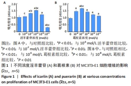
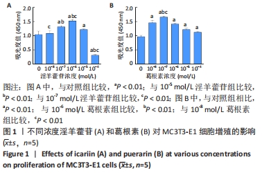
2.1 淫羊藿苷、葛根素对MC3T3-E1最适浓度的选择 2.1.1 细胞增殖浓度选择 采用CCK-8法测定葛根素和淫羊藿苷促进MC3T3-E1细胞增殖的最适浓度,结果显示,淫羊藿苷的最适浓度为10-6 mol/L,葛根素的最适浓度为10-7 mol/L,见图1。"

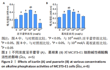
2.1.2 细胞分化浓度选择 采用碱性磷酸酶染色法测定葛根素和淫羊藿苷促进成骨细胞分化的最适浓度,结果显示,淫羊藿苷的最适浓度为10-6 mol/L,葛根素的最适浓度为10-7 mol/L,见图2。"
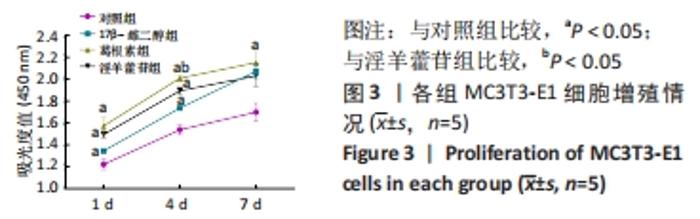
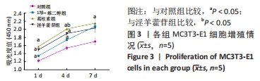
2.2 比较最适葛根素、淫羊藿苷浓度对MC3T3-E1细胞生物学性能的影响 2.2.1 细胞分化浓度选择 如图3所示,在相同时间点,葛根素组和淫羊藿苷组与对照组相比均能促进细胞增殖,且差异有显著性意义(P < 0.05)。葛根素组、淫羊藿苷组、17β-雌二醇组对细胞的增殖促进能力逐渐增加,呈现出时间依赖性。经比较发现,第4天时葛根素组的吸光度值最高,其次为淫羊藿苷组,差异有显著性意义(P < 0.05)。由此判断3种药物中葛根素促进增殖作用最强,其次为淫羊藿苷,17β-雌二醇最弱。"
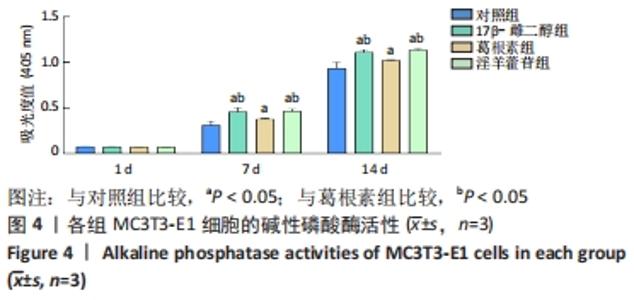
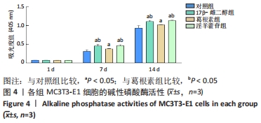
2.2.2 细胞分化能力的比较 淫羊藿苷组在成骨诱导7,14 d时的碱性磷酸酶活性均高于对照组和葛根素组(P < 0.05),见图4,淫羊藿苷组与17β-雌二醇组吸光度值差异无显著性意义,表明淫羊藿苷对细胞分化的促进作用方面优于葛根素。"

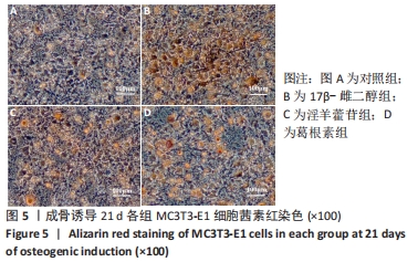
2.2.3 成骨细胞矿化能力 钙沉积水平是矿化的直接标志。光镜下观察及分析计算钙化结节面积,在成骨诱导第21天,4组钙化结节的沉积水平出现显著差异(P < 0.05)。葛根素组、淫羊藿苷组、17β-雌二醇组钙化结节明显多于对照组。17β-雌二醇组钙结节阳性染色面积占比最大,淫羊藿苷组较葛根素组钙化结节面积占比大,差异有显著性意义(P < 0.01),见图5,图6。"
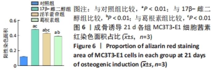
"

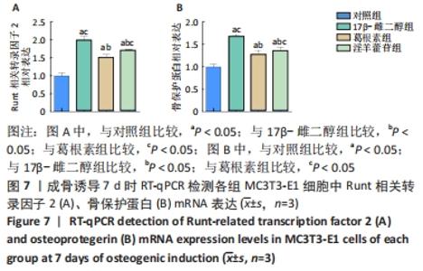
2.2.4 Runt相关转录因子2、骨保护蛋白 mRNA表达 成骨诱导培养第7天时17β-雌二醇组Runt相关转录因子2、骨保护蛋白 mRNA表达量最高,其次为淫羊藿苷组,与葛根素组和对照组相比差异均有显著性意义,葛根素组高于对照组(P < 0.05),见图7。"

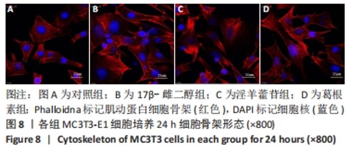
2.2.5 微丝骨架在细胞中的分布 如图8所示,培养24 h,各实验组与对照组相比细胞黏附数量多,细胞铺展面积大。细胞完全伸展,伪足多且清晰;细胞中微丝束朝向细胞长轴方向,大量的微丝结构建立了良好的接触,形成完整网络。"

| [1] EL-RASHIDY AA, ROETHER JA, HARHAUS L, et al. Regenerating bone with bioactive glass scaffolds: A review of in vivo studies in bone defect models. Acta Biomater. 2017;62:1-28. [2] 李祖浩,王辰宇,王中汉.骨质疏松性骨缺损的治疗进展:支架植入与局部药物递送[J].中国组织工程研究,2018,22(18):2939-2945. [3] BUHRMANN C, HONARVAR A, SETAYESHMEHR M, et al. Herbal Remedies as Potential in Cartilage Tissue Engineering: An Overview of New Therapeutic Approaches and Strategies. Molecules. 2020;25(13): 3075. [4] AWALE G, WONG E, RAJPURA K, et al. Engineered Bone Tissue with Naturally-Derived Small Molecules. Curr Pharm Des. 2017;23(24): 3585-3594. [5] WU Y, XIA L, ZHOU Y, et al. Evaluation of osteogenesis and angiogenesis of icariin loaded on micro/nano hybrid structured hydroxyapatite granules as a local drug delivery system for femoral defect repair. J Mater Chem B. 2015;3(24):4871-4883. [6] 刘永庆,李琪佳,甘洪全,等.葛根素联合国产多孔钽对人成骨细胞COL-I、OC、OPN表达和细胞增殖的影响[J].中国骨质疏松杂志, 2017,23(2):236-243. [7] AN J, YANG H, ZHANG Q, et al. Natural products for treatment of osteoporosis: The effects and mechanisms on promoting osteoblast-mediated bone formation. Life Sci. 2016;147:46-58. [8] XU Y, LI L, TANG Y, et al. Icariin promotes osteogenic differentiation by suppressing Notch signaling. Eur J Pharmacol. 2019;865:172794. [9] SUZUKI H, TATEI K, OHSHIMA N, et al. Regulation of MC3T3-E1 differentiation by actin cytoskeleton through lipid mediators reflecting the cell differentiation stage. Biochem Biophys Res Commun. 2019; 514(2):393-400. [10] ZENG X, FENG Q, ZHAO F, et al. Puerarin inhibits TRPM3/miR-204 to promote MC3T3-E1 cells proliferation, differentiation and mineralization. Phytother Res. 2018;32(6):996-1003. [11] WANG Z, WANG D, YANG D, et al. The effect of icariin on bone metabolism and its potential clinical application. Osteoporos Int. 2018; 29(3):535-544. [12] 何罕亮.雌激素对成骨细胞MC3T3-E1的影响及其相关机制研究[D].苏州:苏州大学,2018. [13] CLEMENTINI M, MORLUPI A, AGRESTINI C, et al. Immediate versus delayed positioning of dental implants in guided bone regeneration or onlay graft regenerated areas: a systematic review. Int J Oral Maxillofac Surg. 2013;42(5):643-650. [14] LU J, HAO Y, ZHAO W, et al. Molecular, Cellular and Pharmaceutical Aspects of Autologous Grafts for Peri-implant Hard and Soft Tissue Defects. Curr Pharm Biotechnol. 2017;18(1):85-94. [15] YAMADA Y, HARA K, NAKAMURA S, et al. Minimally invasive approach with tissue engineering for severe alveolar bone atrophy case. Int J Oral Maxillofac Surg. 2013;42(2):260-263. [16] KŘÍŽOVÁ L, DADÁKOVÁ K, KAŠPAROVSKÁ J, et al. Isoflavones. Molecules. 2019;24(6):1076. [17] SIROTKIN AV, HARRATH AH. Phytoestrogens and their effects. Eur J Pharmacol. 2014;741:230-236. [18] WANG Y, WANG WL, XIE WL, et al. Puerarin stimulates proliferation and differentiation and protects against cell death in human osteoblastic MG-63 cells via ER-dependent MEK/ERK and PI3K/Akt activation. Phytomedicine. 2013;20(10):787-796. [19] YANG X, ZHENG H, LIU Y, et al. Puerarin for OVX-Induced Postmenopausal Osteoporosis in Murine Model: Systematic Review and Meta-Analysis. Curr Stem Cell Res Ther. 2020;15(1):37-42. [20] XIE L, LIU N, XIAO Y, et al. In Vitro and In Vivo Osteogenesis Induced by Icariin and Bone Morphogenetic Protein-2: A Dynamic Observation. Front Pharmacol. 2020;11:1058. [21] HO MX, POON CC, WONG KC, et al. Icariin, but Not Genistein, Exerts Osteogenic and Anti-apoptotic Effects in Osteoblastic Cells by Selective Activation of Non-genomic ERα Signaling. Front Pharmacol. 2018;9:474. [22] URASOPON N, HAMADA Y, CHERDSHEWASART W, et al. Preventive effects of Pueraria mirifica on bone loss in ovariectomized rats. Maturitas. 2008;59(2):137-148. [23] TIYASATKULKOVIT W, MALAIVIJITNOND S, CHAROENPHANDHU N, et al. Pueraria mirifica extract and puerarin enhance proliferation and expression of alkaline phosphatase and type I collagen in primary baboon osteoblasts. Phytomedicine. 2014;21(12):1498-1503. [24] HO-SHUI-LING A, BOLANDER J, RUSTOM LE, et al. Bone regeneration strategies: Engineered scaffolds, bioactive molecules and stem cells current stage and future perspectives. Biomaterials. 2018;180:143-162. [25] FENG Q, CHENG SY, YANG R, et al. Puerarin promotes the viability and differentiation of MC3T3-E1 cells by enhancing LC3B-mediated autophagy through downregulation of miR-204. Exp Ther Med. 2020; 19(2):883-890. [26] 吴曦,彭锐.不同浓度淫羊藿苷对人骨髓间充质干细胞成骨分化的影响[J].中国组织工程研究,2018,22(5):669-674. [27] 张姝江,李颖,白波,等.中药骨康方对体外培养大鼠成骨细胞骨架保护作用的初步实验研究[J].世界中医药,2016,11(5):880-883. [28] WEI Q, WANG B, HU H, et al. Icaritin promotes the osteogenesis of bone marrow mesenchymal stem cells via the regulation of sclerostin expression. Int J Mol Med. 2020;45(3):816-824. [29] YANG A, YU C, LU Q, et al. Mechanism of Action of Icariin in Bone Marrow Mesenchymal Stem Cells. Stem Cells Int. 2019;2019:5747298. [30] HONG D, CHEN HX, YU HQ, et al. Morphological and proteomic analysis of early stage of osteoblast differentiation in osteoblastic progenitor cells. Exp Cell Res. 2010;316(14):2291-2300. [31] SHANBHAG S, PANDIS N, MUSTAFA K, et al. Bone tissue engineering in oral peri-implant defects in preclinical in vivo research: A systematic review and meta-analysis. J Tissue Eng Regen Med. 2018;12(1): e336-e349. [32] RISTELI L, RISTELI J. Biochemical markers of bone metabolism. Ann Med. 1993;25(4):385-393. [33] ZHAN XQ, ZENG XW, ZHANG YY, et al. Puerarin promotes the viability and differentiation of MC3T3‑E1 cells by miR‑204‑regulated Runx2 upregulation. Mol Med Rep. 2017;16(5):6262-6268. [34] BOYLE WJ, SIMONET WS, LACEY DL. Osteoclast differentiation and activation. Nature. 2003;423(6937):337-342. |
| [1] | Dang Yi, Du Chengyan, Yao Honglin, Yuan Nenghua, Cao Jin, Xiong Shan, Zhang Dingmei, Wang Xin. Hormonal osteonecrosis and oxidative stress [J]. Chinese Journal of Tissue Engineering Research, 2023, 27(9): 1469-1476. |
| [2] | Liu Xiaolin, Mu Xinyue, Ma Ziyu, Liu Shutai, Wang Wenlong, Han Xiaoqian, Dong Zhiheng. Effect of hydrogel-loaded simvastatin microspheres on osteoblast proliferation and differentiation [J]. Chinese Journal of Tissue Engineering Research, 2023, 27(7): 998-1003. |
| [3] | Liu Wentao, Feng Xingchao, Yang Yi, Bai Shengbin. Effect of M2 macrophage-derived exosomes on osteogenic differentiation of bone marrow mesenchymal stem cells [J]. Chinese Journal of Tissue Engineering Research, 2023, 27(6): 840-845. |
| [4] | Wu Boyu, Ye Kai, Chen Jiahan, Wang Jianghua, Wurikaixi·Aiyiti, Jiang Houfeng, Teng Yong. Biocompatibility of 3D printed polyetheretherketone/hydroxyapatite composites [J]. Chinese Journal of Tissue Engineering Research, 2023, 27(12): 1932-1937. |
| [5] | Wei Yanzhao, Zheng Xiaohan, Gao Shijun, Huang Ting, Wei Xufang, Chen Xinxu, Zhao Zhenqiang. Expression of autocrine macrophage migration inhibitory factor and its receptors of human embryonic stem cells [J]. Chinese Journal of Tissue Engineering Research, 2023, 27(1): 34-41. |
| [6] | Yang Yan, Wang Jingxian, Zhang Ronghong, Li Chen, Fan Anran, Cui Dongbing, Wu Shumei. Effects of conditioned media of different sources on the proliferation of human dental pulp stem cells [J]. Chinese Journal of Tissue Engineering Research, 2023, 27(1): 49-53. |
| [7] | Lan Qian, Gu Yangcong, Xiao Xin, Bi Xueting, Li Na. Human periodontal ligament stem cells-derived exosomes interfere with the proliferation and differentiation of MC3T3-E1 cells [J]. Chinese Journal of Tissue Engineering Research, 2023, 27(1): 54-58. |
| [8] | Liu Wentao, Feng Xingchao, Yang Yi, Bai Shengbin. Effect of M2 macrophage-derived exosomes on osteogenic differentiation of bone marrow mesenchymal stem cells [J]. Chinese Journal of Tissue Engineering Research, 2022, 26(在线): 1-6. |
| [9] | Wang Jing, Xiong Shan, Cao Jin, Feng Linwei, Wang Xin. Role and mechanism of interleukin-3 in bone metabolism [J]. Chinese Journal of Tissue Engineering Research, 2022, 26(8): 1260-1265. |
| [10] | Xiao Hao, Liu Jing, Zhou Jun. Research progress of pulsed electromagnetic field in the treatment of postmenopausal osteoporosis [J]. Chinese Journal of Tissue Engineering Research, 2022, 26(8): 1266-1271. |
| [11] | Zhang Jinglin, Leng Min, Zhu Boheng, Wang Hong. Mechanism and application of stem cell-derived exosomes in promoting diabetic wound healing [J]. Chinese Journal of Tissue Engineering Research, 2022, 26(7): 1113-1118. |
| [12] | Huang Chenwei, Fei Yankang, Zhu Mengmei, Li Penghao, Yu Bing. Important role of glutathione in stemness and regulation of stem cells [J]. Chinese Journal of Tissue Engineering Research, 2022, 26(7): 1119-1124. |
| [13] | Li Jiajun, Xia Tian, Liu Jiamin, Chen Feng, Chen Haote, Zhuo Yinghong, Wu Weifeng. Molecular mechanism by which icariin regulates osteogenic signaling pathways in the treatment of steroid-induced avascular necrosis of the femoral head [J]. Chinese Journal of Tissue Engineering Research, 2022, 26(5): 780-785. |
| [14] | Yang Sidi, Wang Qian, Xu Nuo, Wang Ronghan, Jin Chuanqi, Lu Ying, Dong Ming. Biodentine enhances the proliferation and differentiation of osteoblasts through upregulating bone morphogenetic protein-2 [J]. Chinese Journal of Tissue Engineering Research, 2022, 26(4): 516-520. |
| [15] | Zhou Liang, Chen Xingzhen, Li Zhenyu, Zhang Zekun, Duan Guoqing. The mechanism of lncRNA HOTAIR in interleukin-1beta-mediated osteoarthritis [J]. Chinese Journal of Tissue Engineering Research, 2022, 26(35): 5607-5613. |
| Viewed | ||||||
|
Full text |
|
|||||
|
Abstract |
|
|||||