Chinese Journal of Tissue Engineering Research ›› 2023, Vol. 27 ›› Issue (34): 5448-5454.doi: 10.12307/2023.839
Previous Articles Next Articles
Preparation of cartilage decellularized extracellular matrix-loaded composite nanofiber scaffolds based on two-nozzle electrospinning
Teng Jianxiang1, Zhu Jisheng1, Yuan Daizhu2, Wang Zhen1, Zhou Yuhu2, Tian Xiaobin1, 2
- 1Guizhou Medical University, Guiyang 550001, Guizhou Province, China; 2Department of Orthopedics, Affiliated Hospital of Guizhou Medical University, Guiyang 550004, Guizhou Province, China
-
Received:2022-11-02Accepted:2022-12-15Online:2023-12-08Published:2023-04-20 -
Contact:Tian Xiaobin, Chief physician, Guizhou Medical University, Guiyang 550001, Guizhou Province, China; Department of Orthopedics, Affiliated Hospital of Guizhou Medical University, Guiyang 550004, Guizhou Province, China -
About author:Teng Jianxiang, Master candidate, Physician, Guizhou Medical University, Guiyang 550001, Guizhou Province, China -
Supported by:Guizhou Science and Technology Plan Project, No. [2021]072 (to TXB)
CLC Number:
Cite this article
Teng Jianxiang, Zhu Jisheng, Yuan Daizhu, Wang Zhen, Zhou Yuhu, Tian Xiaobin. Preparation of cartilage decellularized extracellular matrix-loaded composite nanofiber scaffolds based on two-nozzle electrospinning[J]. Chinese Journal of Tissue Engineering Research, 2023, 27(34): 5448-5454.
share this article
Add to citation manager EndNote|Reference Manager|ProCite|BibTeX|RefWorks

2.1 软骨dECM各组分的含量 见图1。"
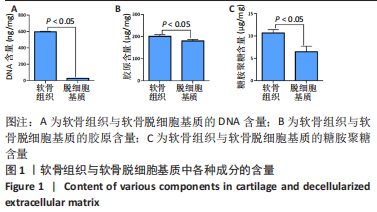

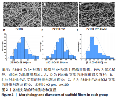
经过酶、化学及超声振荡洗涤处理后,所制备的软骨dECM中残余的DNA含量为(25.53±2.06) ng/mg,较正常软骨组织DNA含量(595.83±6.35) ng/mg明显减少(P < 0.05 ),去除DNA含量约95%。dECM中的胶原含量(179.53±7.20) μg/mg,较软骨组织中的胶原含量(200.43±7.78) μg/mg降低(P < 0.05),保留了胶原约90%;dECM中的糖胺聚糖含量为(6.48±1.23) μg/mg,较正常软骨组织的糖胺聚糖含量(10.69±0.74) μg/mg降低(P < 0.05),保留了糖胺聚糖约60%。 2.2 各组支架物理性能表征结果 2.2.1 各组支架形态学观察 各组支架扫描电镜观察结果如图2所示,各组支架纤维呈随机分布,纤维之间互相连接呈现多孔结构。通过测量各组支架的纤维直径,P34HB支架的纤维直径为(435.00±68.60) nm,P34HB-PVA支架纤维直径为(401.00±54.90) nm,P34HB-PVA-dECM支架的纤维直径为(314.00±58.80) nm。"
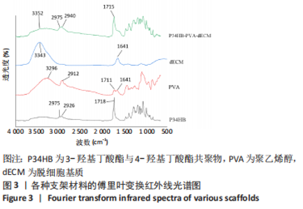
2.2.2 支架成分检测结果 通过傅里叶红外光谱检测显示,所构建的P34HB-PVA-dECM支架在波数为3 352 cm-1处出现宽峰,这归属于dECM与PVA中-OH伸缩振动共同形成的宽峰,2 975,2 940 cm-1处的波峰分别归属于P34HB与PVA中烃基C-H键反对称伸缩振动与对称伸缩振动峰,1 715 cm-1处出现的强峰为C=O键伸缩振动峰,为P34HB的特征峰,见图3。综上,上述3种材料的特征峰均有出现在此红外光谱图中,说明采用双喷头静电纺丝的方法成功地将软骨dECM负载于支架上。"
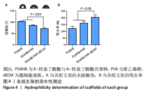
2.2.3 各组支架亲水性能检测结果 P34HB、P34HB-PVA、P34HB-PVA-dECM支架的接触角分别为(102.91±2.21)°,(85.44±2.96)°,(71.42±4.01)°;与其他组相比,P34HB-PVA-dECM支架的水接触角最小(P < 0.05),见图4A。各组支架的吸水率结果如图4B所示,P34HB、P34HB-PVA、P34HB-PVA-dEC支架的吸水率分别为(121.04±15.49)%,(160.38±15.20)%,(203.75±17.99)%,P34HB-PVA-dECM支架的吸水率最高(P < 0.05),说明P34HB-PVA-dECM支架的亲水性最好。"

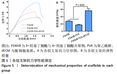
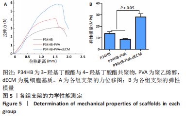
2.2.4 各组支架的力学性能检测结果 各组支架的力-位移曲线如图5A所示,P34HB、P34HB-PVA、P34HB-PVA-dECM支架的弹性模量分别为(13.75±1.65),(8.68±0.78),(28.12±2.81) MPa。 P34HB-PVA-dECM支架的弹性模量大于P34HB、P34HB-PVA支架(P < 0.05),见图5B。说明经过交联剂处理后,P34HB-PVA-dECM支架具有更好的力学性能。"
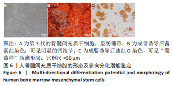
2.3 骨髓间充质干细胞的培养与鉴定 经过传代、纯化,第3代的骨髓间充质干细胞形态如图6A所示,细胞呈纺锤形;经过14 d的成骨诱导后行茜素红染色,光镜下可见明显的钙结节,如图6B所示;经过14 d的成脂诱导后行油红O染色,可见“葡萄样”脂滴形成,如图6C所示。综上所述,所提取的细胞具有多向分化潜能,鉴定为骨髓间充质干细胞。"


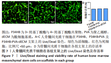
2.4.1 骨髓间充质干细胞在各组支架上的Live/Dead 染色 骨髓间充质干细胞在3组支架上培养3 d以后的Live/Dead 染色结果,如图7A-C所示,可见少量的红色细胞(死细胞),大多数为绿色细胞(活细胞);各组支架上细胞存活率如图7D所示,3组支架上的细胞存活率均超过了95%,说明大多数细胞在3组支架上能存活,3组支架无明显细胞毒性。"
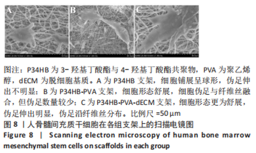
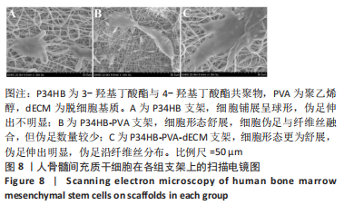
2.4.2 骨髓间充质干细胞在各组支架上的黏附形态 骨髓间充质干细胞在各组支架上培养48 h后的扫描电镜图,如图8所示。扫描电镜观察可见,P34HB支架上的细胞铺展呈球形,伪足伸出不明显;在P34HB-PVA支架上,细胞形态舒展,细胞伪足与纤维丝融合,但伪足数量较少;在P34HB-PVA-dECM支架上,细胞形态舒展更为舒展,伪足伸出明显,伪足沿纤维丝分布,说明细胞与P34HB-PVA-dECM支架结合得更好。"
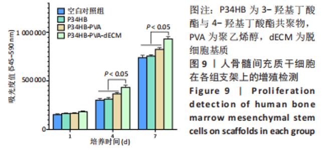
2.4.3 细胞在各组支架上的增殖实验结果 各组支架上骨髓间充质干细胞阿尔玛蓝结果,如图9所示。培养 1 d时,3 组支架上的细胞增殖吸光度比较差异无显著性意义(P > 0.05);培养4,7 d时,P34HB-PVA-dECM支架的的细胞增殖吸光度明显高于P34HB、P34HB-PVA支架(P < 0.05),说明随着培养时间的延长,细胞在P34HB-PVA-dECM支架中的增殖速度最快,dECM的加入促进了细胞的增殖。"

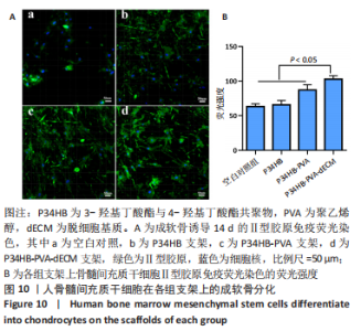
2.4.4 各组支架体外成软骨诱导活性检测结果 骨髓间充质干细胞在各组支架上进行成软骨诱导培养14 d后,Ⅱ型胶原免疫荧光染色结果如图10A所示,各组细胞均出现染色阳性区域,说明骨髓间充质干细胞成功分化为软骨细胞。Image J定量分析结果如图10B所示,P34HB-PVA-dECM支架较其他两支架的荧光强度更强,即分泌的Ⅱ型胶原量更多(P < 0.05),表明P34HB-PVA-dECM支架具有更强的成软骨诱导活性。"


2.5 纳米纤维支架的生物相容性 由支架生物活性表征结果可知,P34HB-PVA-dECM支架具有良好的生物相容性。"

| [1] ZHANG FX, LIU P, DING W, et al. Injectable mussel-Inspired highly adhesive hydrogel with exosomes for endogenous cell recruitment and cartilage defect regeneration. Biomaterials. 2021;278:121169. [2] WANG S, YANG L, CAI B, et al. Injectable hybrid inorganic nanoscaffold as rapid stem cell assembly template for cartilage repair. Natl Sci Rev. 2022; 9(4):nwac037. [3] AL-QURAYSHI Z, WAFA EI, ROSSI MEYER MK, et al. Tissue engineering the pinna: comparison and characterization of human decellularized auricular biological scaffolds. ACS Appl Bio Mater. 2021;4(9):7234-7242. [4] ORTH P, GAO L, MADRY H. Microfracture for cartilage repair in the knee: a systematic review of the contemporary literature. Knee Surg Sport Traumatol Arthrosc. 2020;28(3):670-706. [5] TIAN G, JIANG S, LI J, et al. Cell-free decellularized cartilage extracellular matrix scaffolds combined with interleukin 4 promote osteochondral repair through immunomodulatory macrophages: in vitro and in vivo preclinical study. Acta Biomater. 2021;127:131-145. [6] BLUM JC, SCHENCK TL, BIRT A, et al. Artificial decellularized extracellular matrix improves the regenerative capacity of adipose tissue derived stem cells on 3D printed polycaprolactone scaffolds. J Tissue Eng. 2021;12: 20417314211022242. [7] 胡秋羽,杨龙,杨勇,等.聚己二酸丁二醇酯-对苯二甲酸丁二醇酯/Ⅰ型胶原取向纤维促进前交叉韧带断裂后腱-骨愈合[J].中国组织工程研究,2022,26(27):4314-4319. [8] XUE J, WU T, DAI Y, et al. Electrospinning and electrospun nanofibers: Methods, materials, and applications. Chem Rev. 2019;119(8):5298-5415. [9] BARHOUM A, PAL K, RAHIER H, et al. Nanofibers as new-generation materials: from spinning and nano-spinning fabrication techniques to emerging applications. Appl Mater Today. 2019;(17):1-35. [10] KANIUK Ł, STACHEWICZ U. Development and advantages of biodegradable PHA polymers based on electrospun PHBV fibers for tissue engineering and other biomedical applications. ACS Biomater Sci Eng. 2021;7(12):5339-5362. [11] BASNETT P, MATHARU RK, TAYLOR CS, et al. Harnessing polyhydroxyalkanoates and pressurized gyration for hard and soft tissue engineering. ACS Appl Mater Interfaces. 2021;13:32624-32639. [12] YE C, HU P, MA MX, et al. PHB/PHBHHx scaffolds and human adipose-derived stem cells for cartilage tissue engineering. Biomaterials. 2009;30(26):4401-4406. [13] 刘鋆,杨龙,王伟宇,等.聚 3-羟基丁酸酯 4-羟基丁酸酯/聚乙二醇/氧化石墨烯组织工程支架的制备和性能评价[J].中国组织工程研究,2021,25(22):3466-3472. [14] MA MX, LIU Q, YE C, et al. Preparation of P3HB4HB/(Gelatin + PVA) composite scaffolds by coaxial electrospinning and its biocompatibility evaluation. Biomed Res Int. 2017;(2017):9251806. [15] MONZAVI SM, KAJBAFZADEH AM, SABETKISH S, et al. Extracellular matrix scaffold using decellularized Cartilage for hyaline cartilage regeneration. Adv Exp Med Biol. 2021;1345:209-223. [16] CHEN M, FENG Z, GUO W, et al. PCL-MECM- Based hydrogel hybrid scaffolds and meniscal fibrochondrocytes promote whole meniscus regeneration in a rabbit meniscectomy model. ACS Appl Mater Interfaces. 2019;11(44): 41626-41639. [17] IRANI M, NODEH SM. PVA/kappa-carrageenan/Au/camptothecin/pegylated-polyurethane/paclitaxel nanofibers against lung cancer treatment. RSC Adv. 2022;12(25):16310-16318. [18] YANG L, ZHAO Y, CUI DB, et al. Coaxial bioelectrospinning of P34HB/PVA microfibers biomimetic scaffolds with simultaneity cell-laden for improving bone regeneration. Mater Design. 2022;213:110349. [19] 赵超尘.脱细胞基质覆层壳聚糖纤维载体的制备和生物学评价[D].广州:南方医科大学,2020. [20] LIU HW, SU WT, LIU CY, et al. Highly organized porous gelatin-based scaffold by microfluidic 3D-foaming technology and dynamic culture for cartilage tissue engineering. Int J Mol Sci. 2022;23(15):8449. [21] XI Y, GE J, GUO Y, et al. Biomimetic elastomeric polypeptidebased nanofibrous matrix for overcoming multidrug-resistant bacteria and enhancing full-thickness wound healing/skin regeneration. ACS Nano. 2018; 12:10772-10784. [22] ZELINKA A, ROELOFS AJ, KANDEL RA, et al. Cellular therapy and tissue engineering for cartilage repair. Osteoarthritis Cartilage. 2022;30(12): 1547-1560. [23] ZHANG Q, NETTLESHIP I, SCHMELZER E, et al. Tissue engineering and regenerative medicine therapies for cell senescence in bone and cartilage. Tissue Eng Part B Rev. 2020;26(1):64-78. [24] KIM HS, MANDAKHBAYAR N, KIM HW, et al. Protein-reactive nanofibrils decorated with cartilage-derived decellularized extracellular matrix for osteochondral defects. Biomaterials. 2021;269:120214. [25] HONG J, YEO M, YANG GH, et al. Cell-electrospinning and its application for tissue engineering. Int J Mol Sci. 2019;20:6208. [26] XING H, LEE H, LUO L, et al. Extracellular matrix-derived biomaterials in engineering cell function. Biotechnol Adv. 2020;42:107421. [27] FIORDALISI M, SILVA AJ, BARBOSA M, et al. Decellularized scaffolds for intervertebral disc regeneration. Trends Biotechnol. 2020;38(9):947-951. [28] GAO S, CHEN M, WANG P, et al. An electrospun fiber reinforced scaffold promotes total meniscus regeneration in rabbit meniscectomy model. Acta Biomater. 2018;73:127-140. [29] ZHANG X, CHEN X, HONG H, et al. Decellularized extracellular matrix scaffolds: Recent trends and emerging strategies in tissue engineering. Bioact Mater. 2021;23(10):15-31. [30] ROTHRAUFF BB, COLUCCINO L, GOTTARDI R, et al. Efficacy of thermoresponsive, photocrosslinkable hydrogels derived from decellularized tendon and cartilage extracellular matrix for cartilage tissue engineering. J Tissue Eng Regen Med. 2018;12(1):e159-e170. [31] GAO S, GUO W, CHEN M, et al. Fabrication and characterization of electrospun nanofibers composed of decellularized meniscus extracellular matrix and polycaprolactone for meniscus tissue engineering. J Mater Chem B. 2017;5(12):2273-2285. [32] MISZUK JM, XU T, YAO Q, et al. Functionalization of PCL-3D electrospun nanofibrous scaffolds for improved BMP2-induced bone formation. Appl Mater Today. 2018;10:194-202. [33] 蒲丛丛,何建新,崔世忠,等.电场对双喷头喷气静电纺纳米纤维成形的影响[J].材料导报,2013,27(7):86-89. [34] 任浩征,潘超,许迎,等.ZnO 表面改性 Zn-Cu 组织工程支架的研究[J].表面技术,2021,50(2):58-65. [35] LI X, LIU M, CHEN F, et al. Design of hydroxyapatite bioceramics with micro-/nano-topographies to regulate the osteogenic activities of bone morphogenetic protein-2 and bone marrow stromal cells. Nanoscale. 2020; 12(13):7284-7300. [36] GIRAO AF, SEMITELA A, PEREIRA AL, et al. Microfabrication of a biomimetic arcade-like electrospun scaffold for cartilage tissue engineering applications. J Mater Sci Mater Med. 2020;31(8):69. [37] GRESHAM RCH, BAHNEY CS, LEACH JK. Growth factor delivery using extracellular matrix-mimicking substrates for musculoskeletal tissue engineering and repair. Bioact Mater. 2021;6(7):1945-1956. [38] ZHU W, CAO L, SONG C, et al. Cell-derived decellularized extracellular matrix scaffolds for articular cartilage repair. Int J Artif Organs. 2021;44(4):269-281. [39] SHAHVERDI F, BARATI A, SALEHI E, et al. Biaxial electrospun nanofibers based on chitosan-poly (vinyl alcohol) and poly (Ɛ-caprolactone) modified with CeAlO3 nanoparticles as potential wound dressing materials. Int J Biol Macromol. 2022;221:736-750. [40] PEDRAM RAD Z, MOKHTARI J, ABBASI M, et al. Calendula officinalis extract/PCL/Zein/Gum arabic nanofibrous bio-composite scaffolds via suspension, two-nozzle and multilayer electrospinning for skin tissue engineering. Int J Biol Macromol. 2019;135:530-543. |
| [1] | Sun Kexin, Zeng Jinshi, Li Jia, Jiang Haiyue, Liu Xia. Mechanical stimulation enhances matrix formation of three-dimensional bioprinted cartilage constructs [J]. Chinese Journal of Tissue Engineering Research, 2023, 27(在线): 1-7. |
| [2] | Huang Linke, Wei Linhua, Jiang Jie, Liu Qian, Chen Weiwei. Effects of estrogen combined with treadmill exercise on bone mass and articular cartilage in ovariectomized mice [J]. Chinese Journal of Tissue Engineering Research, 2023, 27(8): 1166-1171. |
| [3] | Xu Xingxing, Wen Chaoju, Meng Maohua, Wang Qinying, Chen Jingqiao, Dong Qiang. Carbon nanomaterials in oral implant [J]. Chinese Journal of Tissue Engineering Research, 2023, 27(7): 1062-1070. |
| [4] | Yang Yitian, Wang Lu, Yao Wei, Zhao Bin. Application of the interaction between biological scaffolds and macrophages in bone regeneration [J]. Chinese Journal of Tissue Engineering Research, 2023, 27(7): 1071-1079. |
| [5] | Li Cheng, Zheng Guoshuang, Kuai Xiandong, Yu Weiting. Alginate scaffold in articular cartilage repair [J]. Chinese Journal of Tissue Engineering Research, 2023, 27(7): 1080-1088. |
| [6] | Xu Cong, Zhao He, Sun Yan. Regeneration of facial nerve injury repaired by biomaterial nerve conduits [J]. Chinese Journal of Tissue Engineering Research, 2023, 27(7): 1089-1095. |
| [7] | Chen Shisong, Liu Xiaohong, Xu Zhiyun. Current status and prospects of bioprosthetic heart valves [J]. Chinese Journal of Tissue Engineering Research, 2023, 27(7): 1096-1102. |
| [8] | Shi Yehong, Wang Cheng, Chen Shijiu. Early thrombosis and prevention of small-diameter blood vessel prosthesis [J]. Chinese Journal of Tissue Engineering Research, 2023, 27(7): 1110-1116. |
| [9] | Tang Haotian, Liao Rongdong, Tian Jing. Application and design of piezoelectric materials for bone defect repair [J]. Chinese Journal of Tissue Engineering Research, 2023, 27(7): 1117-1125. |
| [10] | Xu Yan, Li Ping, Lai Chunhua, Zhu Peijun, Yang Shuo, Xu Shulan. Piezoelectric materials for vascularized bone regeneration [J]. Chinese Journal of Tissue Engineering Research, 2023, 27(7): 1126-1132. |
| [11] | Li Xinyue, Li Xiheng, Mao Tianjiao, Tang Liang, Li Jiang. Three-dimensional culture affects morphology, activity and osteogenic differentiation of human periodontal ligament stem cells [J]. Chinese Journal of Tissue Engineering Research, 2023, 27(6): 846-852. |
| [12] | Li Xiaoyin, Yang Xiaoqing, Chen Shulian, Li Zhengchao, Wang Ziqi, Song Zhen, Zhu Daren, Chen Xuyi. Collagen/silk fibroin scaffold combined with neural stem cells in the treatment of traumatic spinal cord injury [J]. Chinese Journal of Tissue Engineering Research, 2023, 27(6): 890-896. |
| [13] | Yuan Bo, Xie Lide, Fu Xiumei. Schwann cell-derived exosomes promote the repair and regeneration of injured peripheral nerves [J]. Chinese Journal of Tissue Engineering Research, 2023, 27(6): 935-940. |
| [14] | Qin Yuxing, Ren Qiangui, Li Zilong, Quan Jiaxing, Shen Peifeng, Sun Tao, Wang Haoyu. Action mechanism and prospect of bone microvascular endothelial cells for treating femoral head necrosis [J]. Chinese Journal of Tissue Engineering Research, 2023, 27(6): 955-961. |
| [15] | Xiong Bohan, Yu Yang, Lu Xiaojun, Wang Xu, Yang Tengyun, Zhang Yaozhang, Liao Xinyu, Zhou Xiaoxiang, He Lu, Li Yanlin. Research progress in promoting tendon to bone healing during anterior cruciate ligament reconstruction [J]. Chinese Journal of Tissue Engineering Research, 2023, 27(5): 779-786. |
| Viewed | ||||||
|
Full text |
|
|||||
|
Abstract |
|
|||||