Chinese Journal of Tissue Engineering Research ›› 2023, Vol. 27 ›› Issue (33): 5314-5319.doi: 10.12307/2023.748
Previous Articles Next Articles
Lymphatic endothelial cells-derived exosomes promote axonal regeneration following peripheral nerve injury
Huang Jinsheng1, Zhang Geyi1, Li Senrui1, Li Jiangnan1, Lu Laijin2, Zhou Nan1
- 1Department of Orthopedics, First Affiliated Hospital of Zhengzhou University, Zhengzhou 450000, Henan Province, China; 2Department of Hand and Foot Surgery, First Hospital of Jilin University, Changchun 130031, Jilin Province, China
-
Received:2022-10-31Accepted:2022-12-12Online:2023-11-28Published:2023-03-30 -
Contact:Zhou Nan, MD, Associate professor, Department of Orthopedics, First Affiliated Hospital of Zhengzhou University, Zhengzhou 450000, Henan Province, China -
About author:Huang Jinsheng, Master candidate, Department of Orthopedics, First Affiliated Hospital of Zhengzhou University, Zhengzhou 450000, Henan Province, China -
Supported by:National Natural Science Foundation of China, No. 82071388 (to ZN); China Postdoctoral Science Foundation Program, No. 2019M660175 (to ZN); Henan Provincial Excellent Youth Science Foundation, No. 212300410077 (to ZN)
CLC Number:
Cite this article
Huang Jinsheng, Zhang Geyi, Li Senrui, Li Jiangnan, Lu Laijin, Zhou Nan. Lymphatic endothelial cells-derived exosomes promote axonal regeneration following peripheral nerve injury[J]. Chinese Journal of Tissue Engineering Research, 2023, 27(33): 5314-5319.
share this article
Add to citation manager EndNote|Reference Manager|ProCite|BibTeX|RefWorks
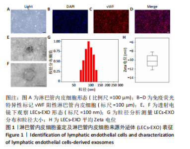
2.1 实验动物数量分析 参加实验的大鼠数量共30只,进入结果分析数量30只,中途无脱落。 2.2 淋巴管内皮细胞鉴定及LECs-EXO表征 显微镜下显示淋巴管内皮细胞呈铺路石状贴壁生长,细胞形态不规则。免疫荧光图像示淋巴管内皮细胞特异性表达内皮细胞标志物vWF。透射电镜下观察LECs-EXO呈典型圆盘状双层膜结构,粒径分析结果示LECs-EXO平均粒径大小为90.06 nm,LECs-EXO平均Zeta电位为-10.21 mV,见图1。"


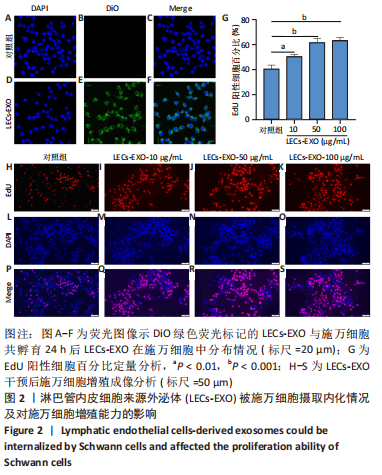
2.3 LECs-EXO细胞摄取及对施万细胞增殖能力的影响 DiO标记LECs-EXO被用于研究施万细胞摄取LECs-EXO效率,DiO标记LECs-EXO与施万细胞孵育24 h后荧光图像示施万细胞胞质中均匀分布绿色荧光信号,表明LECs-EXO被施万细胞高效摄取。EdU细胞增殖实验用于评估LECs-EXO对施万细胞增殖能力的影响,实验结果显示:不同质量浓度(10,50,100 μg/mL)LECs-EXO处理24 h后EdU阳性细胞百分比分别为(50.67±1.53)%,(62.00±3.00)%,(63.67±2.08)%,均高于对照组(40.67±3.06)%,差异有显著性意义(P < 0.05)。上述结果表明,LECs-EXO可增强施万细胞的增殖能力,且这种促增殖能力具有浓度依赖性,当LECs-EXO为50 μg/mL时施万细胞的增殖能力显著增强,见图2。后续动物实验以此浓度继续研究LECs-EXO对周围神经损伤后轴突再生的影响。"
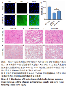
2.4 LECs-EXO坐骨神经标记及对大鼠肌肉湿质量比和神经修复的影响 DiO标记LECs-EXO被用于研究EXO在坐骨神经内分布情况,荧光图像示:术后28 d坐骨神经切片中可见均匀分布绿色荧光,表明LECs-EXO有良好的神经相容性,且能长期存在于坐骨神经中。LECs-EXO治疗组肌肉湿质量比(0.71±0.03)显著高于周围神经损伤组(0.53±0.04),差异有显著性意义(P < 0.05),表明LECs-EXO可抑制坐骨神经损伤后腓肠肌萎缩。坐骨神经苏木精-伊红染色及Masson染色结果显示,LECs-EXO治疗组坐骨神经轴突更加致密,分布均匀且有序;周围神经损伤组坐骨神经轴突排列紊乱无序且可见大量空泡状变性,见图3。"

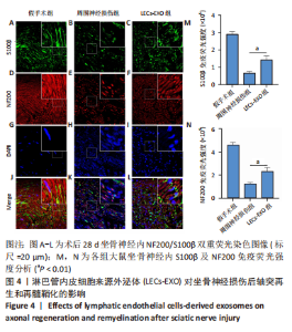
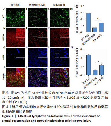
2.5 LECs-EXO对于轴突再生及再髓鞘化的影响 坐骨神经免疫荧光双重染色示:LECs-EXO治疗组NF200及S100β荧光强度(NF200:232 964±31 566;S100β:142 763±21 834)均显著高于周围神经损伤组(NF200:124 809±10 455;S100β:66 461±10 074),差异有显著性意义(P < 0.05),表明LECs-EXO在周围神经损伤后可促进施万细胞增殖及轴突再生,见图4。"
| [1] 陈焱,肖志宏,邢丹谋.周围神经损伤再生与修复的研究进展[J].中华显微外科杂志,2015,38(4):413-416. [2] OLIVER G, KIPNIS J, RANDOLPH GJ, et al. The Lymphatic Vasculature in the 21(st) Century: Novel Functional Roles in Homeostasis and Disease. Cell. 2020;182:270-296. [3] BOUVRÉE K, BRUNET I, DEL TORO R, et al. Semaphorin3A, Neuropilin-1, and PlexinA1 are required for lymphatic valve formation. Circ Res. 2012;111:437-445. [4] FRUEH FS, GOUSOPOULOS E, POWER DM, et al. A potential role of lymphangiogenesis for peripheral nerve injury and regeneration. Med Hypotheses. 2020;135:109470. [5] KALLURI R, LEBLEU VS. The biology, function, and biomedical applications of exosomes. Science. 2020;367(6478):eaau6977. [6] MITTELBRUNN M, SÁNCHEZ-MADRID F. Intercellular communication: diverse structures for exchange of genetic information. Nat Rev Mol Cell Biol. 2012;13:328-335. [7] 袁一鸣,王艳,陈程程,等.外泌体在周围神经损伤进程中的效应[J].中国组织工程研究,2020,24(31):5079-5084. [8] JALKANEN S, SALMI M. Lymphatic endothelial cells of the lymph node. Nat Rev Immunol. 2020;20:566-578. [9] 裴福兴,王光林,林卫.周围神经损伤后免疫反应与神经再生的研究进展[J].中华显微外科杂志,1997,20(2):80-82. [10] PETROVA TV, KOH GY. Biological functions of lymphatic vessels. Science. 2020;369(6500):eaax4063. [11] JACOB L, BOISSERAND LSB, GERALDO LHM, et al. Anatomy and function of the vertebral column lymphatic network in mice. Nat Commun. 2019;10:4594. [12] VOLPI N, AGLIANÒ M, MASSAI L, et al. Lymphatic vessels in human sural nerve: immunohistochemical detection by D2-40. Lymphology. 2006;39:171-173. [13] LIM AH, SULI A, YANIV K, et al. Motoneurons are essential for vascular pathfinding. Development. 2011;138:3847-3857. [14] TAKAMATSU H, TAKEGAHARA N, NAKAGAWA Y, et al. Semaphorins guide the entry of dendritic cells into the lymphatics by activating myosin II. Nat Immunol. 2010;11:594-600. [15] HROMADA C, HARTMANN J, OESTERREICHER J, et al. Occurrence of Lymphangiogenesis in Peripheral Nerve Autografts Contrasts Schwann Cell-Induced Apoptosis of Lymphatic Endothelial Cells In Vitro. Biomolecules. 2022;12(6):820. [16] EKSTRÖM K, VALADI H, SJÖSTRAND M, et al. Characterization of mRNA and microRNA in human mast cell-derived exosomes and their transfer to other mast cells and blood CD34 progenitor cells. J Extracell Vesicles. 2012;1:10. [17] LOPEZ-VERRILLI MA, CAVIEDES A, CABRERA A, et al. Mesenchymal stem cell-derived exosomes from different sources selectively promote neuritic outgrowth. Neuroscience. 2016;320:129-139. [18] LI C, LI X, SHI Z, et al. Exosomes from LPS-preconditioned bone marrow MSCs accelerated peripheral nerve regeneration via M2 macrophage polarization: Involvement of TSG-6/NF-κB/NLRP3 signaling pathway. Exp Neurol. 2022;356:114139. [19] LOPEZ-LEAL R, COURT FA. Schwann Cell Exosomes Mediate Neuron-Glia Communication and Enhance Axonal Regeneration. Cell Mol Neurobiol. 2016;36:429-436. [20] LIU YP, YANG YD, MOU FF, et al. Exosome-Mediated miR-21 Was Involved in the Promotion of Structural and Functional Recovery Effect Produced by Electroacupuncture in Sciatic Nerve Injury. Oxid Med Cell Longev. 2022;2022:7530102. [21] ZHAN C, MA CB, YUAN HM, et al. Macrophage-derived microvesicles promote proliferation and migration of Schwann cell on peripheral nerve repair. Biochem Biophys Res Commun. 2015;468:343-348. [22] GAO B, ZHOU S, SUN C, et al. Brain Endothelial Cell-Derived Exosomes Induce Neuroplasticity in Rats with Ischemia/Reperfusion Injury. ACS Chem Neurosci. 2020;11:2201-2213. [23] ZHANG YZ, LIU F, SONG CG, et al. Exosomes derived from human umbilical vein endothelial cells promote neural stem cell expansion while maintain their stemness in culture. Biochem Biophys Res Commun. 2018;495:892-898. [24] JIANG Y, XIE H, TU W, et al. Exosomes secreted by HUVECs attenuate hypoxia/reoxygenation-induced apoptosis in neural cells by suppressing miR-21-3p. Am J Transl Res. 2018;10:3529-3541. [25] LI R, LI D, WU C, et al. Nerve growth factor activates autophagy in Schwann cells to enhance myelin debris clearance and to expedite nerve regeneration. Theranostics. 2020;10:1649-1677. [26] CARR L, PARKINSON DB, DUN XP. Expression patterns of Slit and Robo family members in adult mouse spinal cord and peripheral nervous system. PLoS One. 2017;12:e0172736. [27] JOHNSTON AP, YUZWA SA, CARR MJ, et al. Dedifferentiated Schwann Cell Precursors Secreting Paracrine Factors Are Required for Regeneration of the Mammalian Digit Tip. Cell Stem Cell. 2016;19:433-448. [28] JONES RE, SALHOTRA A, ROBERTSON KS, et al. Skeletal Stem Cell-Schwann Cell Circuitry in Mandibular Repair. Cell Rep. 2019;28:2757-2766.e5. [29] PARFEJEVS V, DEBBACHE J, SHAKHOVA O, et al. Injury-activated glial cells promote wound healing of the adult skin in mice. Nat Commun. 2018;9:236. [30] MIN Q, PARKINSON DB, DUN XP. Migrating Schwann cells direct axon regeneration within the peripheral nerve bridge. Glia. 2021;69:235-254. [31] HANWRIGHT PJ, QIU C, RATH J, et al. Sustained IGF-1 delivery ameliorates effects of chronic denervation and improves functional recovery after peripheral nerve injury and repair. Biomaterials. 2022; 280:121244. [32] ZOCHODNE DW. The challenges and beauty of peripheral nerve regrowth. J Peripher Nerv Syst. 2012;17:1-18. [33] SCHEIB J, HÖKE A. Advances in peripheral nerve regeneration. Nat Rev Neurol. 2013;9:668-676. [34] JESSEN KR, ARTHUR-FARRAJ P. Repair Schwann cell update: Adaptive reprogramming, EMT, and stemness in regenerating nerves. Glia. 2019; 67:421-437. [35] ARTHUR-FARRAJ PJ, LATOUCHE M, WILTON DK, et al. c-Jun reprograms Schwann cells of injured nerves to generate a repair cell essential for regeneration. Neuron. 2012;75:633-647. [36] JESSEN KR, MIRSKY R, LLOYD AC. Schwann Cells: Development and Role in Nerve Repair. Cold Spring Harb Perspect Biol. 2015;7:a020487. [37] ALLODI I, UDINA E, NAVARRO X. Specificity of peripheral nerve regeneration: interactions at the axon level. Prog Neurobiol. 2012;98: 16-37. [38] LIU T, WANG Y, LU L, et al. SPIONs mediated magnetic actuation promotes nerve regeneration by inducing and maintaining repair-supportive phenotypes in Schwann cells. J Nanobiotechnology. 2022; 20:159. [39] SHEN Y, CHENG Z, CHEN S, et al. Dysregulated miR-29a-3p/PMP22 Modulates Schwann Cell Proliferation and Migration During Peripheral Nerve Regeneration. Mol Neurobiol. 2022;59:1058-1072. [40] SUZUKI T, KADOYA K, ENDO T, et al. Molecular and Regenerative Characterization of Repair and Non-repair Schwann Cells. Cell Mol Neurobiol. 2022;7:12. |
| [1] | Nong Fuxiang, Jiang Zhixiong, Li Yinghao, Xu Wencong, Shi Zhilan, Luo Hui, Zhang Qinglang, Zhong Shuang, Tang Meiwen. Bone cement augmented proximal femoral nail antirotation for type A3.3 intertrochanteric femoral fracturalysis [J]. Chinese Journal of Tissue Engineering Research, 2023, 27(在线): 1-10. |
| [2] | Pan Zhongjie, Qin Zhihong, Zheng Tiejun, Ding Xiaofei, Liao Shijie. Targeting of non-coding RNAs in the pathogenesis of the osteonecrosis of the femoral head [J]. Chinese Journal of Tissue Engineering Research, 2023, 27(9): 1441-1447. |
| [3] | Liu Wentao, Feng Xingchao, Yang Yi, Bai Shengbin. Effect of M2 macrophage-derived exosomes on osteogenic differentiation of bone marrow mesenchymal stem cells [J]. Chinese Journal of Tissue Engineering Research, 2023, 27(6): 840-845. |
| [4] | Li Qicheng, Deng Jin, Fu Xiaoyang, Han Na. Effects of bone marrow mesenchymal stem cells-derived exosomes on hypoxia-treated myoblasts [J]. Chinese Journal of Tissue Engineering Research, 2023, 27(6): 853-859. |
| [5] | Wang Min, Yin Xiushan, Wang Yingxi, Zhang Yan, Zhao Long, Xia Shuyue. Inhalation of bone marrow mesenchymal stem cells-derived exosomes alleviates inflammatory injury in chronic obstructive pulmonary disease [J]. Chinese Journal of Tissue Engineering Research, 2023, 27(6): 827-834. |
| [6] | Zhang Houjun, Deng Bowen, Jiang Shengyuan, Zhao Yi, Ren Jingpei, Xu Lin, Mu Xiaohong. Proteomic analysis of cerebrospinal fluid exosomes derived from cerebral palsy children [J]. Chinese Journal of Tissue Engineering Research, 2023, 27(6): 903-908. |
| [7] | Gao Ting, Ma Xiaohong, Li Xiaorong. Extraction and identification of exosomes from three different sources of ovarian granulosa cells [J]. Chinese Journal of Tissue Engineering Research, 2023, 27(6): 860-865. |
| [8] | Yuan Bo, Xie Lide, Fu Xiumei. Schwann cell-derived exosomes promote the repair and regeneration of injured peripheral nerves [J]. Chinese Journal of Tissue Engineering Research, 2023, 27(6): 935-940. |
| [9] | Xu Qijing, Yang Yichun, Lei Wei, Yang Ying, Yu Jiang, Xia Tingting, Zhang Meng, Zhang Tao, Zhang Qian. Advances and problems in cell-free treatment of diabetic skin chronic wounds [J]. Chinese Journal of Tissue Engineering Research, 2023, 27(6): 962-969. |
| [10] | Chen Guanting, Zhang Linqi, Li Qingru. Meta-analysis of the value of exosomal miRNA for the diagnosis of chronic kidney disease [J]. Chinese Journal of Tissue Engineering Research, 2023, 27(6): 970-976. |
| [11] | Zhou Heshan, Tan Longwang, Liu Chuang, Zhang Chi. Pretreatment methods improve the role of exosomes in spinal cord injury [J]. Chinese Journal of Tissue Engineering Research, 2023, 27(33): 5394-5403. |
| [12] | Wu Zhiwen, Shen Enpu, Li Beibei, Liu Danping, Qi Hui. Exosomes derived from bone marrow mesenchymal stem cells with chondromodulin-1 overexpression affect the proliferation of chondrocytes in osteoarthritis [J]. Chinese Journal of Tissue Engineering Research, 2023, 27(33): 5277-5282. |
| [13] | Wang Yiying, Li Ruiqing, Li Jingwen, Mei Jinjin, Zhang Jianyun, Zhang Lihong, Fan Yongfu, Guo Jian. Exosome-mediated cellular communication: a potential biomarker for Parkinson’s disease [J]. Chinese Journal of Tissue Engineering Research, 2023, 27(24): 3883-3891. |
| [14] | Wang Dongxu, Ren Jing, Li Jiping, Wang Yusu, Hu Pengfei, Zhang Guokun, Li Chunyi. Deer antler stem cell-derived exosomes prevent alcoholic liver injury via modulating nuclear factor kappa B signaling pathway in mice [J]. Chinese Journal of Tissue Engineering Research, 2023, 27(24): 3824-3830. |
| [15] | Zhang Weiye, Zhan Jiawen, Zhu Liguo, Wang Shangquan, Chen Ming, Wei Xu, Feng Minshan, Yu Jie, Han Tao, Cai Chuhao, Zhou Shuaiqi, Shao Chenchen. Effect of nucleus pulposus cells-derived exosomes under cyclic mechanical tension on endplate chondrocytes [J]. Chinese Journal of Tissue Engineering Research, 2023, 27(2): 223-229. |
| Viewed | ||||||
|
Full text |
|
|||||
|
Abstract |
|
|||||