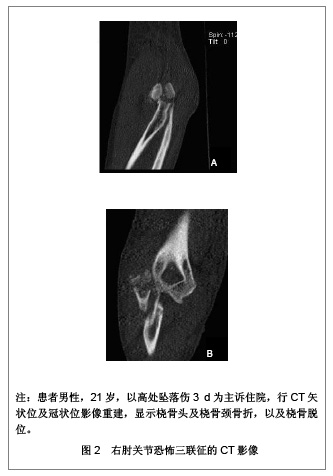
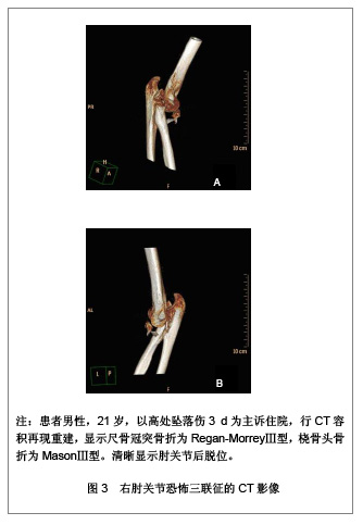
| [1] 王云钊.中华影像医学:骨肌系统卷[M].北京:人民卫生出版社, 2002:128.[2] 陈明祥,邰学祥,陈娟,等.螺旋CT多平面和三维重建在肘关节骨折中的诊断价值[J].实用放射学杂志,2007,23(5):656-657.[3] 李德龙,刘斯平,王振波.16层MSCT不同重建方法在骨与关节疾病诊断中的应用价值研究[J].中国中西医结合影像学杂志,2007, 5(2):106-109.[4] Mulkens TH, Bellinck P, Baeyaert M, et al. Use of an automatic exposure control mechanism for dose optimization in multi-detector row CT examinations: clinical evaluation. Radiology. 2005;237(1):213-223.[5] van Riet RP, Bain GI, Baird R, et al. Simultaneous reconstruction of medial and lateral elbow ligaments for instability using a circumferential graft. Tech Hand Up Extrem Surg. 2006;10(4):239-244.[6] Haapamäki VV, Kiuru MJ, Mustonen AO, et al. Multidetector computed tomography in acute joint fractures. Acta Radiol. 2005;46(6):587-598.[7] Wodecki P, Maiza D, Rozenblum B. Elbow dislocation in children associated with proximal radioulnar translocation. Rev Chir Orthop Reparatrice Appar Mot. 2007;93(2):190-194.[8] 扈延龄,裴国献,李旭,等.三维CT重建对关节内骨折分型术前评价的影响[J].中国矫形外科杂志,2008,16(8):568-570.[9] 何强.MSCT重建技术在肘关节创伤性骨折中的应用[J].实用临床医药杂志,2012,16(13):65-66.[10] 栗占国.类风湿关节炎[M].北京:人民卫生出版社,2009:149.[11] 中国知网.中国学术期刊总库[DB/OL].2013-1-10. https://www.cnki.net[12] 肖观东,周长元,郑超,等.肘关节骨折56例数字化X线摄影分析[J].广东医学,2011,32(6):780-782.[13] 龙响云,方向军,罗祖孝,等.16层螺旋CT多平面重组和容积再现对肘关节损伤的诊断价值[J].重庆医科大学学报,2011,36(8): 985-987.[14] 梁秀梅.MRI对22例肘关节病变的诊断价值[J].重庆医学,2012, 41(10):993-996.[15] Hotchkiss RN. Fractures and dislocations of the elbow. In: Rockwood CA, Green DP, Bucholz RW, et al. Rockwood and Green's fractures in adults. 4th ed.Volume 1. Philadelphia: Lippincott-Raven,1996:929-1024.[16] Ring D, Jupiter JB, Zilberfarb J. Posterior dislocation of the elbow with fractures of the radial head and coronoid. J Bone Joint Surg Am. 2002;84-A(4):547-551.[17] Pugh DM, McKee MD. The "terrible triad" of the elbow. Tech Hand Up Extrem Surg. 2002;6(1):21-29.[18] Amis AA, Miller JH. The mechanisms of elbow fractures: an investigation using impact tests in vitro. Injury. 1995;26(3): 163-168.[19] 张世民.肘关节恐怖三联征的诊治进展[J].同济大学学报(医学版),2010,31(1):5-11.[20] Regan W, Morrey B. Fractures of the coronoid process of the ulna. J Bone Joint Surg Am. 1989;71(9):1348-1354.[21] Mason ML. Some observations on fractures of the head of the radius with a review of one hundred cases. Br J Surg. 1954; 42(172):123-132.[22] Durakbasa MO, Gumussuyu G, Gungor M, et al. Distal humeral coronal plane fractures: management, complications and outcome. J Shoulder Elbow Surg. 2013;22(4):560-566.[23] Riseborough EJ, Radin EL. Intercondylar T fractures of the humerus in the adult. A comparison of operative and non-operative treatment in twenty-nine cases. J Bone Joint Surg Am. 1969;51(1):130-141.[24] Wong AS, Baratz ME. Elbow fractures: distal humerus. J Hand Surg Am. 2009;34(1):176-190.[25] Lumsdaine W, Enninghorst N, Hardy BM, et al. Patterns of CT use and surgical intervention in upper limb periarticular fractures at a level-1 trauma centre. Injury. 2013;44(4): 471-474.[26] McCollough CH, Primak AN, Saba O, et al. Dose performance of a 64-channel dual-source CT scanner. Radiology. 2007;243(3):775-784.[27] Cody DD, Stevens DM, Ginsberg LE. Multi-detector row CT artifacts that mimic disease. Radiology. 2005;236(3):756-761.[28] 王土兴,王立章,俞方荣.多层螺旋CT在骨创伤诊断中的临床价值[J].中国医学影像技术,2003,19(1):118-119.[29] Jelly LM, Evans DR, Easty MJ, et al. Radiography versus spiral CT in the evaluation of cervicothoracic junction injuries in polytrauma patients who have undergone intubation. Radiographics. 2000;20 Spec No:S251-9; discussion S260-262.[30] Klingebiel R, Kentenich M, Bauknecht HC, et al. Comparative evaluation of 64-slice CT angiography and digital subtraction angiography in assessing the cervicocranial vasculature. Vasc Health Risk Manag. 2008;4(4):901-907.[31] Guitton TG, Ring D; Science of Variation Group. Interobserver reliability of radial head fracture classification: two-dimensional compared with three-dimensionalCT. J Bone Joint Surg Am. 2011;93(21):2015-2021.[32] Adams JE, Sanchez-Sotelo J, Kallina CF 4th, et al. Fractures of the coronoid: morphology based upon computer tomography scanning. J Shoulder Elbow Surg. 2012;21(6): 782-788.[33] Kotsianos D, Rock C, Euler E, et al. 3-D imaging with a mobile surgical image enhancement equipment (ISO-C-3D). Initial examples of fracturediagnosis of peripheral joints in comparison with spiral CT and conventional radiography. Unfallchirurg. 2001;104(9):834-838.[34] Rieker O, Mildenberger P, Rudig L, et al. 3D CT of fractures: comparison of volume and surface reconstruction. Rofo. 1998;169(5):490-494.[35] Dewailly M, Rémy-Jardin M, Duhamel A, et al. Computer- aided detection of acute pulmonary embolism with 64-slice multi-detector row computed tomography: impact of the scanning conditions and overall image quality in the detection of peripheral clots. J Comput Assist Tomogr. 2010;34(1): 23-30.[36] Mowatt G, Cummins E, Waugh N, et al. Systematic review of the clinical effectiveness and cost-effectiveness of 64-slice or higher computed tomography angiography as an alternative to invasive coronary angiography in the investigation of coronary artery disease. Health Technol Assess. 2008; 12(17): Iii-iv, ix-143.[37] Gufler H, Schulze CG, Wagner S, et al. MRI for occult physeal fracture detection in children and adolescents. Acta Radiol. 2013.[38] Rhyou IH, Kim KC, Kim KW, et al. Collateral ligament injury in the displaced radial head and neck fracture: correlation with fracture morphology and management strategy to the torn ulnar collateral ligament. J Shoulder Elbow Surg. 2013;22(2): 261-267.[39] Timmerman LA, Schwartz ML, Andrews JR. Preoperative evaluation of the ulnar collateral ligament by magnetic resonance imaging and computed tomography arthrography. Evaluation in 25 baseball players with surgical confirmation. Am J Sports Med. 1994;22(1):26-31; discussion 32.[40] Fritz RC, Steinbach LS, Tirman PF, et al. MR imaging of the elbow. An update. Radiol Clin North Am. 1997;35(1):117-144. |