[1] LANGER R, VACANTI J. Advances in tissue engineering. J Pediatr Surg. 2016;51(1):8-12.
[2] FU X, LIU G, HALIM A, et al. Mesenchymal Stem Cell Migration and Tissue Repair. Cells. 2019;8(8):784.
[3] SHI Y, HU Y, LV C, et al. Effects of Reactive Oxygen Species on Differentiation of Bone Marrow Mesenchymal Stem Cells. Ann Transplant. 2016;21:695-700.
[4] YANG F, LI WB, QU YW, et al. Bone marrow mesenchymal stem cells induce M2 microglia polarization through PDGF-AA/MANF signaling. World J Stem Cells. 2020;12(7):633-658.
[5] YU Y, JIN G, XUE Y, et al. Multifunctions of dual Zn/Mg ion co-implanted titanium on osteogenesis, angiogenesis and bacteria inhibition for dental implants. Acta Biomater. 2017;49:590-603.
[6] NASR AZADANI M, ZAHEDI A, BOWOTO OK, et al. A review of current challenges and prospects of magnesium and its alloy for bone implant applications. Prog Biomater. 2022;11(1):1-26.
[7] WANG J, TANG J, ZHANG P, et al. Surface modification of magnesium alloys developed for bioabsorbable orthopedic implants: a general review. J Biomed Mater Res B Appl Biomater. 2012;100(6):1691-1701.
[8] MOSTAED E, SIKORA-JASINSKA M, DRELICH JW, et al. Zinc-based alloys for degradable vascular stent applications. Acta Biomater. 2018;71:1-23.
[9] LIU Y, RATH B, TINGART M, et al. Role of implants surface modification in osseointegration: A systematic review. J Biomed Mater Res A. 2020; 108(3):470-484.
[10] CHA PR, HAN HS, YANG GF, et al. Biodegradability engineering of biodegradable Mg alloys: tailoring the electrochemical properties and microstructure of constituent phases. Sci Rep. 2013;3:2367.
[11] KIM HK, HAN HS, LEE KS, et al. Comprehensive study on the roles of released ions from biodegradable Mg-5 wt% Ca-1 wt% Zn alloy in bone regeneration. J Tissue Eng Regen Med. 2017;11(10):2710-2724.
[12] ZHANG C, LIN J, NGUYEN NT, et al. Antimicrobial Bioresorbable Mg-Zn-Ca Alloy for Bone Repair in a Comparison Study with Mg-Zn-Sr Alloy and Pure Mg. ACS Biomater Sci Eng. 2020;6(1):517-538.
[13] FARTO-VAAMONDE X, AURIEMMA G, AQUINO RP, et al. Post-manufacture loading of filaments and 3D printed PLA scaffolds with prednisolone and dexamethasone for tissue regeneration applications. Eur J Pharm Biopharm. 2019;141:100-110.
[14] GUO H, LI F, ZHOU YB. The Development of a Biomimetic Nanoscaled Spinal Cage Based on Three-Dimensional Reconstruction Imaging. Biomaterials and Tissue Engineering. 2016;7(6):526-530.
[15] GOSSET A, BARREIRO-VILLAVERDE D, BECERRA PERMUY JC, et al. Experimental and Numerical Investigation of the Extrusion and Deposition Process of a Poly(lactic Acid) Strand with Fused Deposition Modeling. Polymers (Basel). 2020;12(12):2885.
[16] CHEN Q, SHOU P, ZHENG C, et al. Fate decision of mesenchymal stem cells: adipocytes or osteoblasts? Cell Death Differ. 2016;23(7): 1128-1139.
[17] LIU Z, CHANG H, HOU Y, et al. Lentivirus‑mediated microRNA‑26a overexpression in bone mesenchymal stem cells facilitates bone regeneration in bone defects of calvaria in mice. Mol Med Rep. 2018; 18(6):5317-5326.
[18] KHAYAT S, FANAEI H, GHANBARZEHI A. Minerals in Pregnancy and Lactation: A Review Article. J Clin Diagn Res. 2017;11(9):QE01-QE05.
[19] XUE J, SINGH S, ZHOU Y, et al. A biodegradable 3D woven magnesium-based scaffold for orthopedic implants. Biofabrication. 2022;14(3). doi: 10.1088/1758-5090/ac73b8.
[20] ZHANG N, WANG W, ZHANG X, et al. The effect of different coatings on bone response and degradation behavior of porous magnesium-strontium devices in segmental defect regeneration. Bioact Mater. 2020;6(6):1765-1776.
[21] QI T, WENG J, YU F, et al. Insights into the Role of Magnesium Ions in Affecting Osteogenic Differentiation of Mesenchymal Stem Cells. Biol Trace Elem Res. 2021;199(2):559-567.
[22] ABED E, MOREAU R. Importance of melastatin-like transient receptor potential 7 and magnesium in the stimulation of osteoblast proliferation and migration by platelet-derived growth factor. Am J Physiol Cell Physiol. 2009;297(2):C360-C368.
[23] CAI S, LEI T, LI N, et al. Effects of Zn on microstructure, mechanical properties and corrosion behavior of Mg–Zn alloys. Mater Sci Eng C Mater Biol Appl. 2012;32(8):2570-2577.
[24] HE G, WU Y, ZHANG Y, et al. Addition of Zn to the ternary Mg-Ca-Sr alloys significantly improves their antibacterial property. J Mater Chem B. 2015;3(32):6676-6689.
[25] SEONG JW, KIM WJ. Development of biodegradable Mg-Ca alloy sheets with enhanced strength and corrosion properties through the refinement and uniform dispersion of the Mg₂Ca phase by high-ratio differential speed rolling. Acta Biomater. 2015;11:531-542.
[26] YOSHIZAWA S, BROWN A, BARCHOWSKY A, et al. Magnesium ion stimulation of bone marrow stromal cells enhances osteogenic activity, simulating the effect of magnesium alloy degradation. Acta Biomater. 2014;10(6):2834-2842.
[27] WU L, FEYERABEND F, SCHILLING AF, et al. Effects of extracellular magnesium extract on the proliferation and differentiation of human osteoblasts and osteoclasts in coculture. Acta Biomater. 2015;27: 294-304.
[28] ZHANG X, CHEN Q, MAO X. Magnesium Enhances Osteogenesis of BMSCs by Tuning Osteoimmunomodulation. Biomed Res Int. 2019; 2019:7908205.
[29] LIU J, ZENG H, XIAO P, et al. Sustained Release of Magnesium Ions Mediated by a Dynamic Mechanical Hydrogel to Enhance BMSC Proliferation and Differentiation. ACS Omega. 2020;5(38):24477-24486.
[30] WANG Z, LIU Q, LIU C, et al. Mg2+ in β-TCP/Mg-Zn composite enhances the differentiation of human bone marrow stromal cells into osteoblasts through MAPK-regulated Runx2/Osx. J Cell Physiol. 2020;235(6):5182-5191.
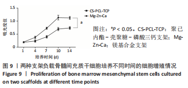
|