[1] MENON DK, SCHWAB K, WRIGHT DW, et al. Position statement: definition of traumatic brain injury. Arch Phys Med Rehabil. 2010;91(11):1637-1640.
[2] GBD 2016 NEUROLOGY COLLABORATORS. Global, regional, and national burden of neurological disorders, 1990-2016: a systematic analysis for the Global Burden of Disease Study 2016. Lancet Neurol. 2019;18(5):459-480.
[3] MAAS AIR, FITZGERALD M, GAO G, et al. Traumatic brain injury over the past 20 years: research and clinical progress. Lancet Neurol. 2022;21(9):768-770.
[4] JIANG JY, GAO GY, FENG JF, et al. Traumatic brain injury in China. Lancet Neurol. 2019;18(3):286-295.
[5] ANCANS J. Cell therapy medicinal product regulatory framework in Europe and its application for MSC-based therapy development. Front Immunol. 2012;3:253.
[6] HOANG DM, PHAM PT, BACH TQ, et al. Stem cell-based therapy for human diseases. Signal Transduct Target Ther. 2022;7(1):272.
[7] MAAS AIR, MENON DK, ADELSON PD, et al. Traumatic brain injury: integrated approaches to improve prevention, clinical care, and research. Lancet Neurol. 2017;16(12):987-1048.
[8] DEWAN MC, RATTANI A, GUPTA S, et al. Estimating the global incidence of traumatic brain injury. J Neurosurg. 2019;130(4):1080-1097.
[9] ISMAIL H, SHAKKOUR Z, TABET M, et al. Traumatic Brain Injury: Oxidative Stress and Novel Anti-Oxidants Such as Mitoquinone and Edaravone. Antioxidants (Basel). 2020;9(10):943.
[10] NAKAO M, INANAGA D, NAGASE K, et al. Characteristic differences of cell sheets composed of mesenchymal stem cells with different tissue origins. Regen Ther. 2019;11:34-40.
[11] ZHAO J, YU G, CAI M, et al. Bibliometric analysis of global scientific activity on umbilical cord mesenchymal stem cells: a swiftly expanding and shifting focus. Stem Cell Res Ther. 2018;9(1):32.
[12] EL OMAR R, BEROUD J, STOLTZ JF, et al. Umbilical cord mesenchymal stem cells: the new gold standard for mesenchymal stem cell-based therapies? Tissue Eng Part B Rev. 2014;20(5):523-544.
[13] SUN W, GREGORY DA, TOMEH MA, et al. Silk Fibroin as a Functional Biomaterial for Tissue Engineering. Int J Mol Sci. 2021;22(3):1499.
[14] BUITRAGO JO, PATEL KD, EL-FIQI A, et al. Silk fibroin/collagen protein hybrid cell-encapsulating hydrogels with tunable gelation and improved physical and biological properties. Acta Biomater. 2018;69:218-233.
[15] TU Y, CHEN C, SUN HT, et al. Combination of temperature-sensitive stem cells and mild hypothermia: a new potential therapy for severe traumatic brain injury. J Neurotrauma. 2012;29(14):2393-2403.
[16] LIU X, ZHANG G, WEI P, et al. Three-dimensional-printed collagen/chitosan/secretome derived from HUCMSCs scaffolds for efficient neural network reconstruction in canines with traumatic brain injury. Regen Biomater. 2022; 9:rbac043.
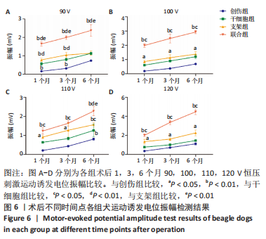
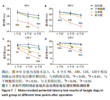
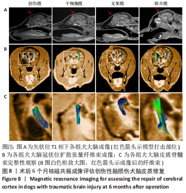
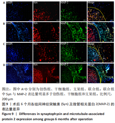
[17] JIANG J, DAI C, NIU X, et al. Establishment of a precise novel brain trauma model in a large animal based on injury of the cerebral motor cortex. J Neurosci Methods. 2018;307:95-105.
[18] CAMERON S, WELTMAN JG, FLETCHER DJ. The prognostic value of admission point-of-care testing and modified Glasgow Coma Scale score in dogs and cats with traumatic brain injuries (2007-2010): 212 cases. J Vet Emerg Crit Care (San Antonio). 2022;32(1):75-82.
[19] ZHU W, CHEN L, WU Z, et al. Bioorthogonal DOPA-NGF activated tissue engineering microunits for recovery from traumatic brain injury by microenvironment regulation. Acta Biomater. 2022;150:67-82.
[20] HSU RS, LI SJ, FANG JH, et al. Wireless charging-mediated angiogenesis and nerve repair by adaptable microporous hydrogels from conductive building blocks. Nat Commun. 2022;13(1):5172.
[21] SUN D, LIU K, LI Y, et al. Intrinsically Bioactive Manganese–Eumelanin Nanocomposites Mediated Antioxidation and Anti‐Neuroinflammation for Targeted Theranostics of Traumatic Brain Injury. Adv Healthc Mater. 2022; 11(16):e2200517.
[22] WEIGHTMAN AP, PICKARD MR, YANG Y, et al. An in vitro spinal cord injury model to screen neuroregenerative materials. Biomaterials. 2014;35(12): 3756-3765.
[23] CHAU AL, GETTY PT, RHODE AR, et al. Superlubricity of pH-responsive hydrogels in extreme environments. Front Chem. 2022;10:891519.
[24] JIANG S, YU Z, ZHANG L, et al. Effects of different aperture-sized type I collagen/silk fibroin scaffolds on the proliferation and differentiation of human dental pulp cells. Regen Biomater. 2021;8(4):rbab028.
[25] MARTÍN-MARTÍN Y, FERNÁNDEZ-GARCÍA L, SANCHEZ-REBATO MH, et al. Evaluation of Neurosecretome from Mesenchymal Stem Cells Encapsulated in Silk Fibroin Hydrogels. Sci Rep. 2019;9(1):8801.
[26] SHAH R, STODULKA P, SKOPALOVA K, et al. Dual Crosslinked Collagen/Chitosan Film for Potential Biomedical Applications. Polymers (Basel). 2019; 11(12):2094.
[27] JIANG X, WU S, KUSS M, et al. 3D printing of multilayered scaffolds for rotator cuff tendon regeneration. Bioact Mater. 2020;5(3):636-643.
[28] MEBARKI M, ABADIE C, LARGHERO J, et al. Human umbilical cord-derived mesenchymal stem/stromal cells: a promising candidate for the development of advanced therapy medicinal products. Stem Cell Res Ther. 2021;12(1):152.
[29] LI J, XU SQ, ZHAO YM, et al. Comparison of the biological characteristics of human mesenchymal stem cells derived from exfoliated deciduous teeth, bone marrow, gingival tissue, and umbilical cord. Mol Med Rep. 2018;18(6):4969-4977.
[30] XIE Q, LIU R, JIANG J, et al. What is the impact of human umbilical cord mesenchymal stem cell transplantation on clinical treatment? Stem Cell Res Ther. 2020;11(1):519.
[31] SUN L, WANG F, CHEN H, et al. Co-Transplantation of Human Umbilical Cord Mesenchymal Stem Cells and Human Neural Stem Cells Improves the Outcome in Rats with Spinal Cord Injury. Cell Transplant. 2019;28(7):893-906.
[32] CAO T, CHEN H, HUANG W, et al. hUC-MSC-mediated recovery of subacute spinal cord injury through enhancing the pivotal subunits β3 and γ2 of the GABAA receptor. Theranostics. 2022;12(7):3057-3078.
[33] CHEN C, HU N, WANG J, et al. Umbilical cord mesenchymal stem cells promote neurological repair after traumatic brain injury through regulating Treg/Th17 balance. Brain Res. 2022;1775:147711.
[34] CUI L, LUO W, JIANG W, et al. Human umbilical cord mesenchymal stem cell-derived exosomes promote neurological function recovery in rat after traumatic brain injury by inhibiting the activation of microglia and astrocyte. Regen Ther. 2022;21:282-287.
[35] OSIER N, DIXON CE. The Controlled Cortical Impact Model of Experimental Brain Trauma: Overview, Research Applications, and Protocol. Methods Mol Biol. 2016;1462:177-192.
[36] JIANG J, LIU X, CHEN H, et al. 3D printing collagen/heparin sulfate scaffolds boost neural network reconstruction and motor function recovery after traumatic brain injury in canine. Biomater Sci. 2020;8(22):6362-6374.
[37] OLSEN A, BABIKIAN T, DENNIS EL, et al. Functional Brain Hyperactivations Are Linked to an Electrophysiological Measure of Slow Interhemispheric Transfer Time after Pediatric Moderate/Severe Traumatic Brain Injury. J Neurotrauma. 2020;37(2):397-409.
[38] SHARMA D, HOLOWAYCHUK MK. Retrospective evaluation of prognostic indicators in dogs with head trauma: 72 cases (January-March 2011). J Vet Emerg Crit Care (San Antonio). 2015;25(5):631-639.
[39] SAENUBOL P, AKATVIPAT A, PLEUMSAMRAN A, et al. Correlation between bispectral index value and modified Glasgow Coma Scale score in dogs with altered level of consciousness. J Vet Emerg Crit Care (San Antonio). 2021;31(1):52-58.
[40] JANG SH, LEE SJ. Corticoreticular Tract in the Human Brain: A Mini Review. Front Neurol. 2019;10:1188.
[41] LI D, JIAO YM, WANG L, et al. Surgical outcome of motor deficits and neurological status in brainstem cavernous malformations based on preoperative diffusion tensor imaging: a prospective randomized clinical trial. J Neurosurg. 2018;130(1):286-301.
[42] JANG SH, KIM SH, KWON YH. Extensive traumatic axonal injury of brain due to violence: A case report. Medicine (Baltimore). 2018;97(48): e13315.
[43] DOUGLAS DB, RO T, TOFFOLI T, et al. Neuroimaging of Traumatic Brain Injury. Med Sci (Basel). 2018;7(1):2.
[44] LACALLE-AURIOLES M, CASSEL DE CAMPS C, ZORCA CE, et al. Applying hiPSCs and Biomaterials Towards an Understanding and Treatment of Traumatic Brain Injury. Front Cell Neurosci. 2020;14:594304.
[45] LV B, ZHANG X, YUAN J, et al. Biomaterial-supported MSC transplantation enhances cell-cell communication for spinal cord injury. Stem Cell Res Ther. 2021;12(1):36.
[46] MONTICELLI S, NATOLI G. Short-term memory of danger signals and environmental stimuli in immune cells. Nat Immunol. 2013;14(8): 777-784.
[47] ZHU D, JIANG Z, LI N, et al. Insights into the use of genetically modified decellularized biomaterials for tissue engineering and regenerative medicine. Adv Drug Deliv Rev. 2022;188:114413. |