[1] SHARMA L. Osteoarthritis of the Knee. N Engl J Med. 2021;384(1):51-59.
[2] LATOURTE A, KLOPPENBURG M, RICHETTE P. Emerging pharmaceutical therapies for osteoarthritis. Nat Rev Rheumatol. 2020;16(12):673-688.
[3] YUNUS MHM, NORDIN A, KAMAL H. Pathophysiological Perspective of Osteoarthritis. Medicina (Kaunas). 2020;56(11):614.
[4] KATZ JN, ARANT KR, LOESER RF. Diagnosis and Treatment of Hip and Knee Osteoarthritis: A Review. JAMA. 2021;325(6):568-578.
[5] MAHESHWER B, POLCE EM, PAUL K, et al. Regenerative Potential of Mesenchymal Stem Cells for the Treatment of Knee Osteoarthritis and Chondral Defects: A Systematic Review and Meta-analysis. Arthroscopy. 2021;37(1):362-378.
[6] DOYLE EC, WRAGG NM, WILSON SL. Intraarticular injection of bone marrow-derived mesenchymal stem cells enhances regeneration in knee osteoarthritis. Knee Surg Sports Traumatol Arthrosc. 2020; 28(12):3827-3842.
[7] MA W, LIU C, WANG S, et al. Efficacy and safety of intra-articular injection of mesenchymal stem cells in the treatment of knee osteoarthritis: A systematic review and meta-analysis. Medicine (Baltimore). 2020;99(49):e23343.
[8] GUPTA A, CADY C, FAUSER AM, et al. Cell-free Stem Cell-Derived Extract Formulation for Regenerative Medicine Applications. Int J Mol Sci. 2020;21(24):9364.
[9] QIU C, GE Z, CUI W, et al. Human Amniotic Epithelial Stem Cells: A Promising Seed Cell for Clinical Applications. Int J Mol Sci. 2020;21(20): 7730.
[10] ZHANG Q, LAI D. Application of human amniotic epithelial cells in regenerative medicine: a systematic review. Stem Cell Res Ther. 2020; 11(1):439.
[11] LIU QW, HUANG QM, WU HY, et al. Characteristics and Therapeutic Potential of Human Amnion-Derived Stem Cells. Int J Mol Sci. 2021; 22(2):970.
[12] MIKI T. Stem cell characteristics and the therapeutic potential of amniotic epithelial cells. Am J Reprod Immunol. 2018;80(4):e13003.
[13] MUTTINI A, BARBONI B, VALBONETTI L, et al. Amniotic Epithelial Stem Cells: Salient Features and Possible Therapeutic Role. Sports Med Arthrosc Rev. 2018;26(2):70-74.
[14] LEBRETON F, LAVALLARD V, BELLOFATTO K, et al. Insulin-producing organoids engineered from islet and amniotic epithelial cells to treat diabetes. Nat Commun. 2019;10(1):4491.
[15] XU H, ZHANG J, TSANG KS, et al. Therapeutic Potential of Human Amniotic Epithelial Cells on Injuries and Disorders in the Central Nervous System. Stem Cells Int. 2019;2019:5432301.
[16] REN Y, CHEN Y, ZHENG X, et al. Human amniotic epithelial cells ameliorate kidney damage in ischemia-reperfusion mouse model of acute kidney injury. Stem Cell Res Ther. 2020;11(1):410.
[17] SABBAH DA, HAJJO R, SWEIDAN K. Review on Epidermal Growth Factor Receptor (EGFR) Structure, Signaling Pathways, Interactions, and Recent Updates of EGFR Inhibitors. Curr Top Med Chem. 2020;20(10):815-834.
[18] SCHNEIDER MR, SIBILIA M, ERBEN RG. The EGFR network in bone biology and pathology. Trends Endocrinol Metab. 2009;20(10):517-524.
[19] BYRNE PO, HRISTOVA K, LEAHY DJ. EGFR forms ligand-independent oligomers that are distinct from the active state. J Biol Chem. 2020; 295(38):13353-13362.
[20] APPLETON CT, USMANI SE, MORT JS, et al. Rho/ROCK and MEK/ERK activation by transforming growth factor-alpha induces articular cartilage degradation. Lab Invest. 2010;90(1):20-30.
[21] 袁功武,兰生辉,刘曦明.表皮生长因子受体信号通路对骨折愈合的生物学作用[J].中国矫形外科杂志,2018,26(4):328-332.
[22] 袁功武,兰生辉,曾文波,等.吉非替尼对骨折愈合中I、II及X 型胶原基因表达的影响[J].中国矫形外科杂志,2018,26(10):939-944.
[23] 程潭.降钙素对大鼠膝关节骨性关节炎潜在保护作用及其机制[D].石家庄:河北医科大学,2012.
[24] ALIMIRAH F, PULIDO T, VALDOVINOS A, et al. Cellular Senescence Promotes Skin Carcinogenesis through p38MAPK and p44/42MAPK Signaling. Cancer Res. 2020;80(17):3606-3619.
[25] PAN L, NI H, JIN W, et al. Inhibition of ERK or Akt ameliorates intimal hyperplasia via up-regulation of Cx37 and down-regulation of Cx43 in balloon injury rat model. Cardiovasc Diagn Ther. 2020;10(4):658-666.
[26] WU Q, FANG T, LANG H, et al. Comparison of the proliferation, migration and angiogenic properties of human amniotic epithelial and mesenchymal stem cells and their effects on endothelial cells. Int J Mol Med. 2017;39(4):918-926.
[27] MA XF, MA XB, QIAN WJ, et al. Co-Culture of Adipose-Derived Stem Cells and Chondrocytes With Transforming Growth Factor-Beta 3 Promotes Chondrogenic Differentiation. J Craniofac Surg. 2020;31(8): 2355-2359.
[28] KAWAKAMI T, KOMATSU T, YOKOYAMA K, et al. Establishment of co-culture of human induced pluripotent stem cell-derived melanocytes and keratinocytes in vitro. J Dermatol. 2021;48(1):123-125.
[29] HUNTER DJ, MARCH L, CHEW M. Osteoarthritis in 2020 and beyond: a Lancet Commission. Lancet. 2020;396(10264):1711-1712.
[30] KULKARNI P, MARTSON A, VIDYA R, et al. Pathophysiological landscape of osteoarthritis. Adv Clin Chem. 2021;100:37-90.
[31] KALAMEGAM G, MEMIC A, BUDD E, et al. A Comprehensive Review of Stem Cells for Cartilage Regeneration in Osteoarthritis. Adv Exp Med Biol. 2018;1089:23-36.
[32] DÍAZ-PRADO S, MUIÑOS-LÓPEZ E, HERMIDA-GÓMEZ T, et al. Human amniotic membrane as an alternative source of stem cells for regenerative medicine. Differentiation. 2011;81(3):162-171.
[33] MANUELPILLAI U, MOODLEY Y, BORLONGAN CV, et al. Amniotic membrane and amniotic cells: potential therapeutic tools to combat tissue inflammation and fibrosis? Placenta. 2011;32 Suppl 4:S320-325.
[34] LIU XY, CHEN J, ZHOU Q, et al. In vitro tissue engineering of lamellar cornea using human amniotic epithelial cells and rabbit cornea stroma. Int J Ophthalmol. 2013;6(4):425-429.
[35] YANG PJ, YUAN WX, LIU J, et al. Biological characterization of human amniotic epithelial cells in a serum-free system and their safety evaluation. Acta Pharmacol Sin. 2018;39(8):1305-1316.
[36] SANKAR V, MUTHUSAMY R. Role of human amniotic epithelial cell transplantation in spinal cord injury repair research. Neuroscience. 2003;118(1):11-17.
[37] YUGE I, TAKUMI Y, KOYABU K, et al. Transplanted human amniotic epithelial cells express connexin 26 and Na-K-adenosine triphosphatase in the inner ear. Transplantation. 2004;77(9):1452-1454.
[38] HORI J, WANG M, KAMIYA K, et al. Immunological characteristics of amniotic epithelium. Cornea. 2006;25(10 Suppl 1):S53-58.
[39] MIKI T, LEHMANN T, CAI H, et al. Stem cell characteristics of amniotic epithelial cells. Stem Cells. 2005;23(10):1549-1559.
[40] SCHNEIDER MR, WOLF E. The epidermal growth factor receptor ligands at a glance. J Cell Physiol. 2009;218(3):460-466.
[41] ZHANG X, ZHU J, LIU F, et al. Reduced EGFR signaling enhances cartilage destruction in a mouse osteoarthritis model. Bone Res. 2014;2:14015.
[42] ROMANO R, BUCCI C. Role of EGFR in the Nervous System. Cells. 2020; 9(8):1887.
[43] ZHU M, QIN YC, GAO CQ, et al. l-Glutamate drives porcine intestinal epithelial renewal by increasing stem cell activity via upregulation of the EGFR-ERK-mTORC1 pathway. Food Funct. 2020;11(3):2714-2724.
[44] LINDER M, HECKING M, GLITZNER E, et al. EGFR controls bone development by negatively regulating mTOR-signaling during osteoblast differentiation. Cell Death Differ. 2018;25(6):1094-1106.
[45] 任科伟,范卫民,姜雪峰,等.周期性机械应力通过激活EGFR-ERK1/2信号通路促进大鼠软骨细胞增殖和细胞外基质合成[J].江苏医药,2013,39(21):2526-2528.
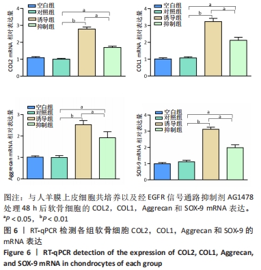
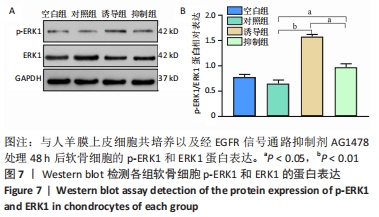
|