[1] KOLASINSKI SL, NEOGI T, HOCHBERG MC, et al. 2019 American College of Rheumatology/Arthritis Foundation Guideline for the Management of Osteoarthritis of the Hand, Hip, and Knee. Arthritis Care Res (Hoboken). 2020;72(2):149-162.
[2] 张莹莹,李旭东,杨佳娟,等.中国40岁及以上人群骨关节炎患病率的Meta分析[J].中国循证医学杂志,2021,21(4):407-414.
[3] 奚阳,石银朋,宋志伟,等.骨关节炎影像学研究进展[J].中国实用内科杂志, 2020,40(2):170-173.
[4] YOON YJ, CHANG S, KIM OY, et al. Three-dimensional imaging of hepatic sinusoids in mice using synchrotron radiation micro-computed tomography. PLoS One. 2013; 8(7):e68600.
[5] RADAKOVICH LB, MAROLF AJ, SHANNON JP, et al. Development of a microcomputed tomography scoring system to characterize disease progression in the Hartley guinea pig model of spontaneous osteoarthritis. Connect Tissue Res. 2018;59(6):523-533.
[6] 张永亮,宓轶群,刚嘉鸿,等.温针灸对膝骨关节炎大鼠关节软骨及形态的影响[J].中国针灸,2016,36(2):175-179.
[7] 杨波,周明旺,吉星,等.中药有效成分调节线粒体保护骨关节炎软骨的研究进展[J].中草药,2021,52(7):2117-2133.
[8] 贾艳辉,卢世璧,汪爱媛,等.CT增强软骨成像技术的研究进展[J].中国医药生物技术,2013,8(2):143-146.
[9] 巫旭娜,许碧莲,田佳,等.用Micro-CT评价大鼠骨质疏松合并骨性关节炎模型[J].中国老年学杂志,2020,40(5):1044-1047.
[10] FLYNN C, HURTIG M, LINDEN AZ. Anionic Contrast-Enhanced MicroCT Imaging Correlates with Biochemical and Histological Evaluations of Osteoarthritic Articular Cartilage. Cartilage. 2020:1947603520924748.
[11] 王江雪,高玉,李辉,等.离子造影剂增强Micro-CT扫描对大鼠关节软骨形态的定量分析[J].生物医学工程研究,2014,33(3):157-161.
[12] DAS NEVES BORGES P, FORTE AE, VINCENT TL, et al. Rapid, automated imaging of mouse articular cartilage by microCT for early detection of osteoarthritis and finite element modelling of joint mechanics. Osteoarthritis Cartilage. 2014; 22(10):1419-1428.
[13] CLARK JN, GARBOUT A, FERREIRA SA, et al. Propagation phase-contrast micro-computed tomography allows laboratory-based three-dimensional imaging of articular cartilage down to the cellular level. Osteoarthritis Cartilage. 2020;28(1):102-111.
[14] KÜN-DARBOIS JD, MANERO F, RONY L, et al. Contrast enhancement with uranyl acetate allows quantitative analysis of the articular cartilage by microCT: Application to mandibular condyles in the BTX rat model of disuse. Micron.2017;97:35-40.
[15] 刘进,傅明,黄广鑫,等.Micro-CT软骨成像监测发育性髋关节发育不良大鼠髋关节软骨早期退变的应用价值[J].中华关节外科杂志(电子版),2013,7(2):213-219.
[16] 刘成磊,郗艳,左后东,等.同步辐射显微CT的人关节软骨三维成像研究[J].CT理论与应用研究,2015,24(6):793-799.
[17] MOHR A, HEISS C, BERGMANN I, et al. Value of micro-CT as an investigative tool for osteochondritis dissecans: A preliminary study with comparison to histology. Acta Radiologica. 2003;44:5. doi:10.1080/j.1600-0455.2003.00113.
[18] 王前源,刘水涛,卫小春,等.软骨下骨在骨性关节炎进程中的作用[J].中国医学前沿杂志(电子版),2017,9(2):53-57.
[19] MOUNTCASTLE SE, ALLEN P, MELLORS BOL, et al. Dynamic viscoelastic characterisation of human osteochondral tissue: understanding the effect of the cartilage-bone interface. BMC Musculoskelet Disord. 2019; 20(1):575.
[20] 常亮,秦江辉,史冬泉,等.骨关节炎与软骨下骨研究进展[J].中华骨与关节外科杂志,2019,12(10):827-832.
[21] POURAN B, ARBABI V, BLEYS RL, et al. Solute transport at the interface of cartilage and subchondral bone plate: Effect of micro-architecture. J Biomech. 2017;52:148-154.
[22] 郭洁梅,陈鹏,肖艳,等.软骨下骨重塑与骨关节炎综述[J].福建中医药,2021, 52(1):54-57.
[23] 陈文杰,李益军,郑小飞,等.软骨下骨的病理改变及其在骨关节炎发病机制中的作用[J].中国骨科临床与基础研究杂志,2020, 12(4):234-241.
[24] 郇松玮,査振刚,王华军,等.miR-214在小鼠骨关节炎软骨及软骨下骨中的表达分析[J].中国矫形外科杂志,2019,27(7):641-645.
[25] PUCHA KA, MCKINNEY JM, FULLER JM, et al. Characterization of OA development between sexes in the rat medial meniscal transection model. Osteoarthr Cartilage Open. 2020;(3):100066.
[26] 耿佳,星月,胡扬帆,等.同步辐射X线显微断层成像在兔膝骨关节炎软骨及软骨下骨三维成像中的应用研究[J].诊断学理论与实践,2020,19(3):238-242.
[27] 谭启钊,牛国栋,赵振达,等.关节软骨与软骨下骨改变在不同骨关节炎动物模型中的特点[J].中国实验动物学报,2019,27(4):450-455.
[28] 孙光华,廖源,彭婷,等.依降钙素对骨质疏松-骨关节炎大鼠的影响[J].中国矫形外科杂志,2020,28(18):1685-1689.
[29] 陶剑锋,王莹,王超,等.豚鼠不同年龄自发性骨关节炎显微变化的观察[J].骨科临床与研究杂志,2019,4(3):167-172.
[30] HOLZER LA ,KRAIGER M, TALAKIC E, et al. Microstructural analysis of subchondral bone in knee osteoarthritis. Osteoporos Int. 2020;31(10):2037-2045. .
[31] 韩广弢,李皓桓,张宇标,等.滑膜在炎性关节病中的作用[J].广西医学,2019, 41(12):1545-1548.
[32] 刘金富,曾平,农焦,等.整合多组微阵列芯片分析骨关节炎患者滑膜中生物标志物和治疗靶点[J].中国组织工程研究,2021,25(23): 3690-3696.
[33] SCANZELLO CR, GOLDRING SR. The role of synovitis in osteoarthritis pathogenesis. Bone. 2012;51(2):249-257.
[34] 郑洁,袁普卫.骨性关节炎的代谢机制研究进展[J].中国骨质疏松杂志,2018, 24(3):406-410.
[35] 宋珊,胡方媛,乔军,等.基于生物信息学途径认识骨关节炎滑膜的生物学标志物[J].中国组织工程研究,2021,25(5):785-790.
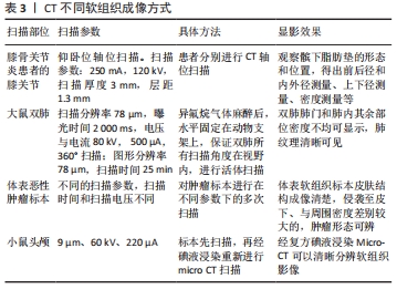
[36] 王琳,张洁,刘太运,等.膝关节骨性关节炎髌下脂肪垫在中药治疗前后的CT变化研究[J].中国中西医结合影像学杂志,2014,12(4): 351-354.
[37] 安国亮,李小丽,王炎,等.Micro CT对骨髓间充质干细胞拮抗矽肺大鼠肺纤维化的效果评价[J].首都医科大学学报,2017,38(2):238-243.
[38] 姜伟乾,陈犹白,陶然,等.显微计算机断层扫描对体表恶性肿瘤成像的研究[J].中华整形外科杂志,2020(3):242-243-244-245-246-247-248-249-250.
[39] 杨榕,李庆祥,王逸飞,等.碘液浸染在Micro-CT下识别小鼠颅底-颞下区肿瘤组织中的应用[J].北京大学学报(医学版),2021,53(3): 598-601.
[40] 江攀,李大鹏,毛良浩,等.滑膜在骨关节炎发病机制及治疗中的作用[J].中国矫形外科杂志,2020,28(5):430-434.
[41] 李晗,陈百成,邵德成,等.血管病理与骨关节炎发病关系的研究进展[J].中国矫形外科杂志,2010,18(1):46-48.
[42] 卢键森,柳鑫,曾春,等.H-型血管在骨关节炎软骨下骨中的表达及作用[J].中国组织工程研究,2017,21(20):3135-3140.
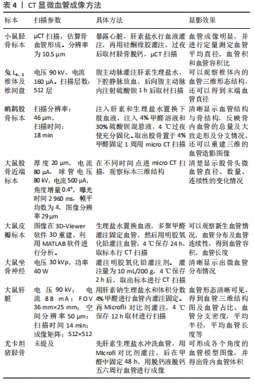
[43] 丁文鸽,戴力扬,蒋雷生.卵巢切除小鼠胫骨干骺端血管的显微CT观察[J].上海交通大学学报(医学版),2008(10):1238-1241.
[44] 徐鸿明,胡斐,王雍立,等.兔椎体软骨终板内血管三维影像结构研究[J].中国脊柱脊髓杂志,2015,25(11):1013-1017.
[45] 范猛,汪爱媛,王玉,等.基于Micro-CT的骨内微血管显影和三维重建[J].南开大学学报(自然科学版),2011,44(1):78-84.
[46] 朱觉新,黄连芳.骨内微血管显影及三维重建在激素致大鼠股骨头缺血性坏死模型中的应用[J].广东医科大学学报,2020,38(5):562-565.
[47] 李国栋,徐永清,何晓清,等.Tempol对大鼠任意皮瓣成活影响的实验研究[J].中国修复重建外科杂志,2016,30(10):1264-1269.
[48] 朱昭炜,毛以华,何波,等.SD大鼠坐骨神经微血管三维可视化研究初探[J].中国修复重建外科杂志,2013,27(2):189-192.
[49] 刘巧遇,李若坤.基于Micro-CT成像对肝癌原位移植瘤血管三维结构的定量研究[J].肝脏,2020,25(12):1290-1293.
[50] KOTSOUGIANI D, HUNDEPOOL CA, BULSTRA LF, et al. Bone vascularized composite allotransplantation model in swine tibial defect: Evaluation of surgical angiogenesis and transplant viability. Microsurgery. 2019; 39(2):160-166.
[51] KLINE TL, ZAMIR M, RITMAN EL. Accuracy of microvascular measurements obtained from micro-CT images. Ann Biomed Eng. 2010;38(9):2851-64.
[52] 许云腾,许丽梅,李慧,等.从筋骨的力学特性探讨膝关节软骨-软骨下骨稳态失衡的生物力学机制[J].风湿病与关节炎,2019,8(12): 43-45+57.
[53] 赵泽明,张柳.NF-κB信号通路与骨关节炎的关系研究进展[J].华北理工大学学报(医学版),2021,23(3):232-238.
|
