中国组织工程研究 ›› 2022, Vol. 26 ›› Issue (9): 1329-1333.doi: 10.12307/2022.423

• 骨科植入物Orthopedic implants • 上一篇    下一篇

夹板外固定与散巴布剂对桡骨骨折模型兔骨愈合的影响

李  睿1,2,史  文3,杨士彩4,吕林蔚1,2,张春秋1,2   

  1. 天津理工大学,1天津市先进机电系统设计与智能控制重点实验室,2机电工程国家级实验教学示范中心,天津市   300384;3嘉斯特华剑医疗器材(天津)有限公司,天津市   300190;4天津中医药大学第二附属医院,天津市   300150
  • 收稿日期:2021-05-18 修回日期:2021-05-19 接受日期:2021-07-14 出版日期:2022-03-28 发布日期:2021-12-09
  • 通讯作者: 吕林蔚,讲师,天津理工大学,天津市先进机电系统设计与智能控制重点实验室,机电工程国家级实验教学示范中心,天津市 300384
  • 作者简介:李睿,男,1996年生,甘肃省金昌市人,汉族,天津理工大学在读硕士,主要从事骨愈合、骨整合研究。
  • 基金资助:
    国家自然科学基金面上项目(12072235),项目负责人:张春秋;天津市自然科学基金项目(18JCZDJC36100),项目负责人:张春秋;天津市科技计划项目生物医学工程科技重大专项(18ZXSGSY00010),项目参与者:张春秋

Effect of splintage and Shenxiaosan cataplasm on fracture healing in rabbits with radial fracture model

Li Rui1,2, Shi Wen3, Yang Shicai4, Lü Linwei1,2, Zhang Chunqiu1,2   

  1. 1Tianjin Key Laboratory for Advanced Mechatronic System Design and Intelligent Control, 2National Experimental Teaching Demonstration Center of Mechanical and Electrical Engineering, Tianjin University of Technology, Tianjin 300384, China; 3Just Huajian Medical Devices (Tianjin) Co., Ltd., Tianjin 300190, China; 4Second Affiliated Hospital of Tianjin University of Traditional Chinese Medicine, Tianjin 300150, China
  • Received:2021-05-18 Revised:2021-05-19 Accepted:2021-07-14 Online:2022-03-28 Published:2021-12-09
  • Contact: Lü Linwei, Lecturer, Tianjin Key Laboratory for Advanced Mechatronic System Design and Intelligent Control, and National Experimental Teaching Demonstration Center of Mechanical and Electrical Engineering, Tianjin University of Technology, Tianjin 300384, China
  • About author:Li Rui, Master candidate, Tianjin Key Laboratory for Advanced Mechatronic System Design and Intelligent Control, and National Experimental Teaching Demonstration Center of Mechanical and Electrical Engineering, Tianjin University of Technology, Tianjin 300384, China
  • Supported by:
    the National Natural Science Foundation of China, No. 12072235 (to ZCQ); Tianjin Natural Science Foundation Project, No. 18JCZDJC36100 (to ZCQ); Science and Technology Major Special Project of Biomedical Engineering of Tianjin Science and Technology Plan Project, No. 18ZXSGSY00010 (to ZCQ)

摘要:

文题释义:
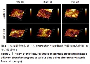
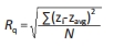
原子力显微镜:可以在大气和液体环境下对各种材料、样品进行纳米区域的物理性质(包括形貌)探测,或者直接进行纳米操纵。实验利用原子力显微镜观测骨痂区域骨组织的颗粒度与表面粗糙度,从而评价骨重建的状态和新生骨组织的力学性能。
三七总皂苷:是三七的主要有效成分,是根据提取、分离技术从优质三七中提取有效的药用成分,包括20多种皂苷活性物质。现代药理学研究表明,三七总皂苷可以促进成骨、抑制骨吸收,是促进创伤骨折疾病痊愈的有效药物。

背景:目前临床治疗长骨骨折的主要手段是石膏或夹板外固定及接骨板或者髓内钉等内固定,同时也可服用加速骨折愈合的药物或者在患处外敷药物。
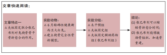
目的:探讨夹板外固定联合散巴布剂对于桡骨骨干骨折的恢复是否有改善作用。
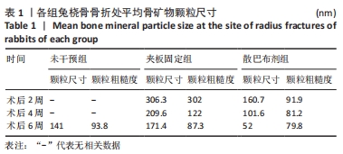
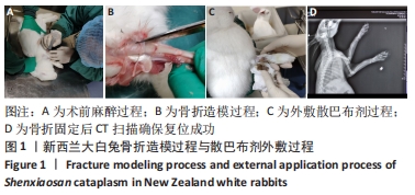
方法:取18只新西兰大白兔,制作左侧桡骨完全性骨折模型,分成3组,未干预组不进行任何干预,夹板固定组用夹板外固定处理,散巴布剂组使用夹板外固定处理并辅助散巴布剂药物外敷(每天换药一次),每组6只。术后2,4,6周,取骨痂位置制备样品,应用原子力显微镜检测骨痂粗糙度和颗粒度。
结果与结论:①夹板固定组、散巴布剂组可以明显观察到骨痂的粗糙表面和明显的颗粒结构,这些矿物颗粒附着在胶原纤维上,呈现出鱼鳞状,其中散巴布剂组的胶原结构远远小于夹板固定组;②随着愈合时间的增长,骨痂矿物颗粒尺寸逐渐减小,散巴布剂组同期骨矿化颗粒尺寸较夹板固定组小47.5%-69.6%;同时随着愈合时间的推移骨痂粗糙度逐渐降低,散巴布剂组术后2周的骨痂粗糙度较夹板固定组小约69%,术后6周两组骨痂粗糙度趋于近似;③结果表明散巴布剂对骨折愈合具有促进作用。

https://orcid.org/0000-0003-0126-8802 (李睿)

中国组织工程研究杂志出版内容重点:人工关节;骨植入物;脊柱;骨折;内固定;数字化骨科;组织工程

关键词: 散巴布剂, 夹板, 骨折愈合, 原子力显微镜, 粗糙度, 颗粒度

Abstract: BACKGROUND: At present, the main clinical methods for the treatment of long bone fractures were external fixation with plaster or splint, and internal fixation such as bone plate or intramedullary nail, simultaneously taking drugs to accelerate fracture healing or applying drugs to the affected area.  
OBJECTIVE: To investigate whether splintage combined with Shenxiaosan cataplasm could improve the rehabilitation of radial shaft fracture.
METHODS:  Eighteen New Zealand white rabbits were used to make a complete fracture model of the left radius, and divided into three groups. No intervention was conducted in the non-intervention group. Models in the splintage group were fixed with splintage. Models in the splintage adjuvant Shenxiaosan group received external fixation with splintage, and adjuvant external application of Shenxiaosan cataplasm (change dressing once a day) (n=6 per group). At 2, 4 and 6 weeks after surgery, the callus position was taken to prepare the sample, and the roughness and particle of callus were measured by atomic force microscope.  
RESULTS AND CONCLUSION: (1) In the splintage group and splintage adjuvant Shenxiaosan group, the rough surface and obvious granular structure of the callus could be clearly observed. These mineral particles were attached to the collagen fibers and presented fish scales. The collagen structure of the splintage adjuvant Shenxiaosan group was much smaller than splintage group. (2) As the healing time increased, the size of bone callus mineral particles gradually decreased. The size of bone mineralization particles in the splintage adjuvant Shenxiaosan group during the same period was 47.5%-69.6% smaller than that of the splintage group. At the same time, as the healing time went on, the roughness of the callus gradually decreased. The roughness of the callus in the splintage adjuvant Shenxiaosan group 2 weeks after the operation was about 69% smaller than that of the splintage group, and the roughness of the callus in the two groups tended to be similar at 6 weeks after the operation. (3) It is indicated that Shenxiaosan cataplasm had a promoting effect on fracture healing.

Key words: Shenxiaosan cataplasm, splintage, fracture healing, atomic force microscope, roughness, granularity

中图分类号: