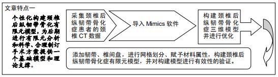
|
[1] MATSUYAMA Y, KAWAKAMI N, YANASE M, et al. Cervical Myelopathy Due to OPLL.J Spinal Disord Tech. 2004;17(5): 401-404.
[2] LEE CH, JAHNG TA, HYUN SJ, et al. Expansive laminoplasty versus laminectomy alone versus laminectomy and fusion for cervical ossification of the posterior longitudinal ligament.J Spinal Disord Tech.2016;29(1):E9-E15.
[3] TSUKIMATO H. A case report autopsy of compression of spinal cord owing to ossification within spinal canal of the cervical spine.Nippon Geka Hokan.1960;29:1003-1007.
[4] KODA M, MOCHIZUKI M, KONISHI H, et al. Comparison of clinical outcomes between laminoplasty, posterior decompression with instrumented fusion, and anterior decompression with fusion for K-line (–) cervical ossification of the posterior longitudinal ligament.Eur Spine J.2016;25(7): 2294-2301.
[5] KAWAGUCHI Y, NAKANO M, YASUDA T, et al. Clinical impact of ossification of the posterior longitudinal ligament progression after cervical laminoplasty.Clin Spine Surg. 2019; 32(3): E133-E139.
[6] BROLIN K, HALLDIN P. Development of a finite element model of the upper cervical spine and a parameter study of ligament characteristics.Spine.2004;29(4):376-385.
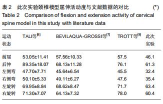
[7] BEVILAQUA-GROSSI D, GONÇALVES MC, CARVALHO GF, et al. Additional Effects of a Physical Therapy Protocol on Headache Frequency, Pressure Pain Threshold, and Improvement Perception in Patients With Migraine and Associated Neck Pain: A Randomized Controlled Trial.Arch Phys Med Rehabil.2016;97(6):866-874.
[8] TALI D, MENAHEM I, VERED E, et al. Upper cervical mobility, posture and myofascial trigger points in subjects with episodic migraine: Case-control study.J Bodyw Mov Ther. 2014;18(4): 569-575.
[9] TROTT PH, PEARCH MJ, RUSTON SA, et al. Three-dimensional analysis of active cervical motion: the effect of age and gender.Clin Biomech .1996;11(4):201-206.
[10] TERAYAMA K, MARUYAMA S, MIYASHITA R, et al. On the ossification of ligament longitudinal posterior in the cervical spine.Seikeigeka.1964;15:1083-1095.
[11] BOODY BS, LENDNER M, VACCARO AR. Ossification of the posterior longitudinal ligament in the cervical spine: a review. Int orthop.2019;43(4):797-805.
[12] KATSUMI K, WATANABE K, IZUMI T, et al. Natural history of the ossification of cervical posterior longitudinal ligament: a three dimensional analysis.Int orthop.2018;42(4):835-842.
[13] YANG H, SUN J, SHI J, et al. Anterior controllable antedisplacement fusion (ACAF) for severe cervical ossification of the posterior longitudinal ligament: comparison with anterior cervical corpectomy with fusion (ACCF).World Neurosurg.2018;115:e428-e436.
[14] KATSUMI K, IZUMI T, ITO T, et al. Posterior instrumented fusion suppresses the progression of ossification of the posterior longitudinal ligament: a comparison of laminoplasty with and without instrumented fusion by three-dimensional analysis. Eur Spine J.2016;25(5):1634-1640.
[15] BONO C, MIN W. Avoiding complications in patients with ankylosing spondylitis undergoing spine surgery.Curr Opin Orthop.2005;16(16):178-183.
[16] ZHAO X, CHEN C, ZHOU T, et al. Analysis of single cage position in transforaminal lumbar interbody fusion through digital images.Int Orthop.2018;42(5):1-7.
|