中国组织工程研究 ›› 2020, Vol. 24 ›› Issue (34): 5467-5472.doi: 10.3969/j.issn.2095-4344.2878

• 组织工程骨材料Tissue-engineered bone • 上一篇    下一篇

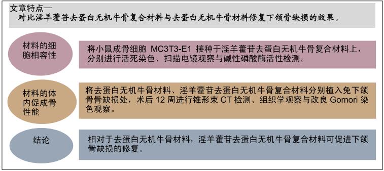
淫羊藿苷去蛋白无机牛骨复合材料与去蛋白无机牛骨材料修复下颌骨缺损

董文杰,张诗扬,赵  磊,王禹锟   

  1. 牡丹江市医学院第二附属医院口腔科,黑龙江省牡丹江市 157000

  • 收稿日期:2019-12-20 修回日期:2019-12-25 接受日期:2020-02-12 出版日期:2020-11-08 发布日期:2020-09-11
  • 通讯作者: 王禹锟,硕士,副主任医师,牡丹江医学院附属第二医院,黑龙江省牡丹江市 157000
  • 作者简介:董文杰,女,1975年生,河北省苏路县人,汉族,副主任医师,主要从事颌面外科方面的研究。
  • 基金资助:
    牡丹江市科学技术计划项目(Z2011s007)

Icariin deproteinized inorganic bovine bone composite and deproteinized inorganic bovine bone material in repairing mandibular defects

Dong Wenjie, Zhang Shiyang, Zhao Lei, Wang Yukun   

  1. Department of Stomatology, Second Affiliated Hospital of Mudanjiang Medical College, Mudanjiang 157000, Heilongjiang Province, China

  • Received:2019-12-20 Revised:2019-12-25 Accepted:2020-02-12 Online:2020-11-08 Published:2020-09-11
  • Contact: Wang Yukun, Master, Associate chief physician, Department of Stomatology, Second Affiliated Hospital of Mudanjiang Medical College, Mudanjiang 157000, Heilongjiang Province, China
  • About author:Dong Wenjie, Associate chief physician, Department of Stomatology, Second Affiliated Hospital of Mudanjiang Medical College, Mudanjiang 157000, Heilongjiang Province, China
  • Supported by:

    the Science and Technology Plan Project of Mudanjiang City, No. Z2011s007

摘要:


文题释义:

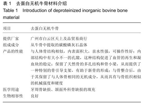
去蛋白无机牛骨:是采用化学提纯法从牛骨中提取的碳酸磷灰石晶体,去除了蛋白及其他的有机成分,而保留了多孔天然骨的无机机构,与人体骨的结构相似,目前已被广泛应用于口腔临床骨再生手术。

淫羊藿苷:是一种具有补肾壮阳、祛风除湿功效的中药,临床多用于免疫力低下、性功能障碍与抗衰老食疗等。淫羊藿苷是淫羊藿的有效药理成分,近年来的研究发现其具有防治骨质疏松,并且其可促进间充质干细胞、成骨细胞的成骨分化与成血管因子的表达。

背景:去蛋白无机牛骨与人体骨的结构相似,目前已被广泛应用于口腔临床骨再生手术,但其缺乏成骨诱导能力。近年来的研究发现淫羊藿具有防治骨质疏松的作用。

目的:观察淫羊藿苷去蛋白无机牛骨复合材料修复下颌骨缺损的效果。
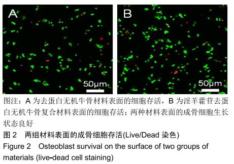
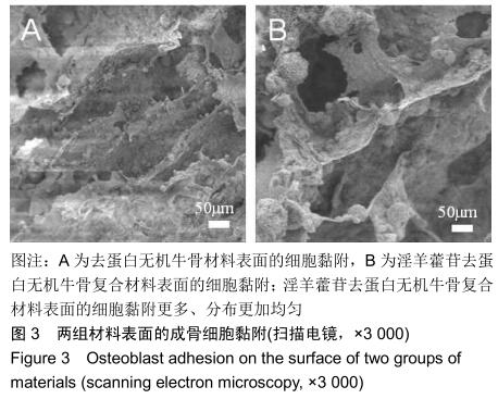
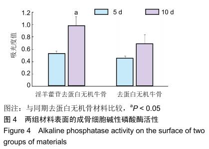
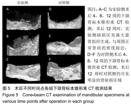
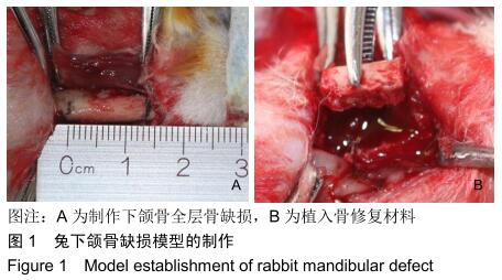
方法将小鼠成骨细胞MC3T3-E1分别接种于淫羊藿苷去蛋白无机牛骨复合材料(观察组)与去蛋白无机牛骨材料(对照组)上,培养7 d后,活死染色观察材料表面细胞的存活,扫描电镜观察材料表面细胞的黏附;培养510 d后,检测细胞分泌碱性磷酸酶情况。在30只新西兰大白兔双侧下颌骨制作13 mm×6 mm×4 mm的全层骨缺损,右侧植入淫羊藿苷去蛋白无机牛骨复合材料(实验侧),左侧植入去蛋白无机牛骨材料(对照侧),术后4812周获取双侧下颌骨组织,分别进行锥形束CT检测、组织学观察与改良Gomori染色观察。实验获得牡丹江医学院实验动物中心伦理委员会批准。
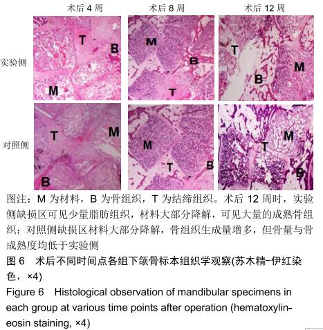
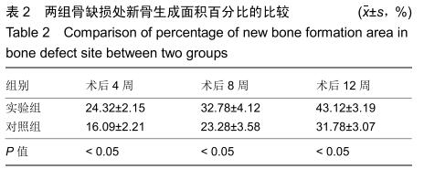
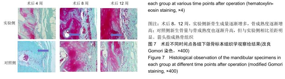
结果与结论①活死染色显示,两组材料表面的成骨细胞生长状态良好;②扫描电镜显示,成骨细胞可在两种材料表面黏附,在观察组材料表面的黏附数量更多、分布更加均匀;③观察组培养10 d的细胞碱性磷酸酶活性高于对照组(P < 0.05);④锥形束CT显示,实验侧骨缺损至术后12周时基本愈合,对照侧术后12周时仍可见骨缺损;⑤术后12周组织学观察显示,实验侧缺损区可见大量成熟骨组织,仅见少量残余材料,可见少量脂肪组织;对照侧虽然材料部分降解,可见较多的新生骨组织,骨成熟度低于实验侧;⑥术后12周改良Gomori染色显示,实验侧可见大量成熟度较高的新生骨组织,对照侧也可见较多成熟度较高的骨组织,但骨量与骨成熟度均不及实验侧;⑦结果表明相对于去蛋白无机牛骨材料,淫羊藿苷去蛋白无机牛骨复合材料可促进下颌骨缺损的修复。

ORCID: 0000-0001-5823-7228(董文杰)

中国组织工程研究杂志出版内容重点:生物材料;骨生物材料; 口腔生物材料; 纳米材料; 缓释材料; 材料相容性;组织工程


关键词: 骨, 材料,  下颌骨,  去蛋白无机牛骨,  淫羊藿苷,  骨缺损,  骨再生

Abstract:

BACKGROUND: Deproteinized inorganic bovine bone is similar to human bone in structure. It has been widely used in oral bone regeneration surgery, but it lacks osteogenic induction ability. In recent years, it has been found that icariin has the effect on preventing and treating osteoporosis.

OBJECTIVE: To observe the effect of icariin deproteinized inorganic bovine bone composite in the repair of mandibular defects.

METHODS: MC3T3-E1 mouse osteoblasts were inoculated on icariin deproteinized inorganic bovine bone composite (observation group) and deproteinized inorganic bovine bone material (control group) respectively. After 7 days of culture, the survival of the cells on the surface of the material was observed by live-dead cell staining. The adhesion of the cells on the surface of the material was observed by scanning electron microscopy. After 5 and 10 days of culture, the alkaline phosphatase activity of the cells was detected. A 13 mm×6 mm×4 mm full-thickness defect of mandible was made in 30 New Zealand white rabbits. The experimental side (right) was implanted with icariin deproteinized inorganic bovine bone composite, and the control side (left) was implanted with deproteinized inorganic bovine bone material. The bilateral mandible tissues were obtained at 4, 8, and 12 weeks after the operation. The cone beam CT detection, histological observation and modified Gomori staining were performed respectively. The experiment was approved by the Ethics Committee of Experimental Animal Center of Mudanjiang Medical College.

RESULTS AND CONCLUSION: (1) Live-dead cell staining demonstrated that the osteoblasts on the surface of the two groups grew well. (2) Scanning electron microscopy showed that osteoblasts adhered to the surfaces of the two materials. The cells on the surface of observation group had more adhesion and more uniform distribution than those in the control group. (3) The alkaline phosphatase activity of observation group cultured for 10 days was higher than that of control group (P < 0.05). (4) Cone-beam CT showed that the bone defect on the experimental side healed at 12 weeks after operation, while the bone defect on the control side could be seen at 12 weeks after operation. (5) Histological observation showed that at 12 weeks after operation, a large number of mature bone tissue could be seen in the defect area on the experimental side, and only a small amount of residual material and adipose tissue could be seen. On the control side, although the material was partially degraded and more new bone tissue could be seen, but the bone maturity was lower than that on the experimental side. (6) Modified Gomori staining showed that at 12 weeks after operation, a large number of new bone tissue with higher maturity could be seen on the experimental side, and more mature bone tissue could be seen on the control side, but the bone mass and maturity were not as good as those on the experimental side. (7) Results confirmed that compared with deproteinized inorganic bovine bone material, icariin deproteinized inorganic bovine bone composite can promote the repair of mandibular defects.

Key words: bone,  material,  mandible,  deproteinized inorganic bovine bone,  icariin,  bone defect,  bone regeneration

中图分类号: