中国组织工程研究 ›› 2020, Vol. 24 ›› Issue (16): 2572-2577.doi: 10.3969/j.issn.2095-4344.1889

• 材料生物相容性 material biocompatibility • 上一篇    下一篇

环保型生物组织样本制备套装在HER2蛋白2+浸润性乳腺癌荧光原位杂交检测中的应用

邱晓阳,王媛媛,刘春鹏,陈洪才,吴  璇,詹晓芬   

  1. 汕头市中心医院病理科,广东省汕头市  515031
  • 收稿日期:2019-05-05 修回日期:2019-05-16 接受日期:2019-06-27 出版日期:2020-06-08 发布日期:2020-03-26
  • 通讯作者: 吴璇,副主任技师,汕头市中心医院病理科,广东省汕头市 515031
  • 作者简介:邱晓阳,女,1979年生,广东省汕头市人,汉族,主管技师,主要从事病理学技术研究。

Application of environment-friendly bio-tissue sample preparation kit in fluorescence in situ hybridization detection of HER2 protein 2-positive invasive breast cancer 

Qiu Xiaoyang, Wang Yuanyuan, Liu Chunpeng, Chen Hongcai, Wu Xuan, Zhan Xiaofen   

  1. Department of Pathology, Shantou Central Hospital, Shantou 515031, Guangdong Province, China
  • Received:2019-05-05 Revised:2019-05-16 Accepted:2019-06-27 Online:2020-06-08 Published:2020-03-26
  • Contact: Wu Xuan, Associate chief technician, Department of Pathology, Shantou Central Hospital, Shantou 515031, Guangdong Province, China
  • About author:Qiu Xiaoyang, Technician in charge, Department of Pathology, Shantou Central Hospital, Shantou 515031, Guangdong Province, China

摘要:
文题释义:

环保型生物组织样本制备套装:无醛、无苯,用于组织样本制备过程中对组织样本的固定、脱水和透明处理,其中固定液是利用无水乙醇、甲醇等化学试剂的蛋白质凝固作用,终止或减少分解酶的作用,防止自溶,保存组织、细胞的离体前结构状态;脱水液是利用能够与水互溶的有机溶液,将已固定和水洗过的组织中的水分彻底驱除;透明液是采用既能与脱水剂互溶,又能作为石蜡溶媒的有机溶剂,使浸蜡过程中石蜡渗入组织中。由于透明剂作用之后其折射指数与组织蛋白折射指数接近,组织显示出半透明状态,因此通常又称此过程为透明。

HER2蛋白:定位于染色体17q12-21.32上,编码相对分子质量为185 000的跨膜受体样蛋白,具有酪氨酸激酶活性。检测方法有免疫组织化学、荧光原位杂交等。HER2是指重要的乳腺癌及胃癌预后判断因子,HER2阳性(过表达或扩增)的乳腺癌/胃癌,其临床特点和生物学行为有特殊表现,治疗模式也与其他类型的乳腺癌/胃癌有很大的区别。目前已有针对该蛋白过度表达的药物——注射用曲妥珠单抗。

背景:HER2状态评估是浸润性乳腺癌治疗及预后重要的生物学指标。固定、脱水、透明和脱蜡等组织前期处理是制作病理石蜡切片后进行HER2蛋白和基因检测的必备程序,也是影响免疫组织化学和荧光原位杂交的重要因素。

目的:探究环保型生物组织样本制备套装在HER2蛋白2+浸润性乳腺癌荧光原位杂交检测中的应用价值。

方法:收集2015年1月至2019年3月汕头市中心医院送检的402例浸润性乳腺癌标本,同一标本对半剖开,使用随机数字表随机分为2组,对照组采用传统试剂甲醛固定-乙醇脱水-二甲苯透明脱蜡进行组织前期处理,制作石蜡切片;实验组采用环保型生物组织样本制备套装(含环保型固定液、脱水液、透明液、脱蜡液)制作切片。采用免疫组织化学法检测两组标本的HER2蛋白表达,进一步对HER2蛋白结果为2+的131例浸润性乳腺癌标本使用荧光原位杂交法检测HER2基因扩增。

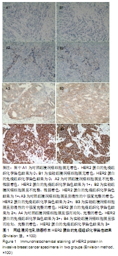
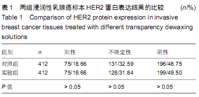
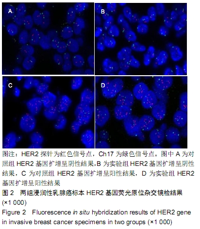
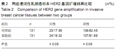
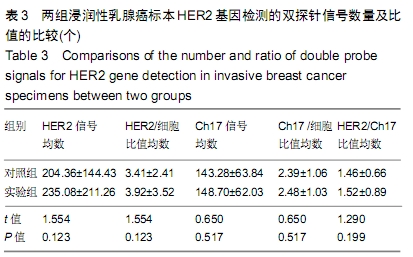
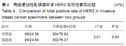
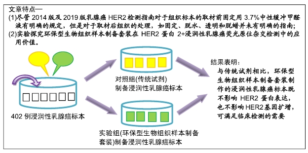
结果与结论:①两组HER2蛋白表达均为特异性的细胞膜着色、细胞定位正确;②两组HER2蛋白阳性率、不确定性率、阴性率比较差异均无显著性意义(P > 0.05),两组HER2蛋白表达符合率为99.00%;③两组HER2基因均背景清晰,HER2和Ch17双探针信号清晰可见,无交差反应、双探针信号精准定位于癌细胞核内;④两组HER2基因均杂交成功,两组杂交成功细胞数量比较差异无显著性意义(P > 0.05);⑤两组HER2基因扩增阳性率、阴性率比较差异均无显著性意义(P > 0.05),两组HER2基因扩增符合率为97.71%;⑥两组HER2基因信号均数、HER2/细胞比值均数、Ch17信号均数、Ch17 /细胞比值均数、HER2/ Ch17比值均数比较差异均无显著性意义(P > 0.05);⑦两组HER2总阳性率比较差异均无显著性意义(P > 0.05);⑧结果表明与传统试剂相比,环保型生物组织样本制备套装制作的浸润性乳腺癌标本既不影响HER2蛋白的表达,也不影响HER2基因扩增,可满足临床检测的需要。

ORCID: 0000-0003-0730-5467(邱晓阳)

中国组织工程研究杂志出版内容重点:生物材料;骨生物材料; 口腔生物材料; 纳米材料; 缓释材料; 材料相容性;组织工程

关键词: 浸润性乳腺癌, 人类表皮生长因子受体2, 免疫组织化学, 荧光原位杂交, 扩增, 固定, 脱水, 透明

Abstract:

BACKGROUND: HER2 status assessment is an important biological index for the treatment and prognosis of invasive breast cancer. Pre-treatment of tissues such as immobilization, dehydration, transparency and dewaxing is a necessary procedure for HER2 protein and gene detection after pathological paraffin sections, and also an important factor affecting immunohistochemistry and fluorescence in situ hybridization.

OBJECTIVE: To explore the application value of environment-friendly biological tissue sample preparation kit in fluorescence in situ hybridization detection of HER2 protein 2-positive invasive breast cancer.

METHODS: 402 invasive breast cancer specimens were collected from Shantou Central Hospital from January 2015 to March 2019. The same specimens were semi-dissected and randomly divided into two groups. The control group was treated by traditional reagent formaldehyde immobilization-ethanol dehydration-xylene transparency and dewaxing, and paraffin sections were made. The experimental group was treated with formaldehyde immobilization-ethanol dehydration-xylene transparent dewaxing. Environment-friendly biological tissue sample preparation kit (including environmentally friendly stationary fluid, dehydration fluid, transparent liquid, dewaxing fluid) was used to make slices. The expression of HER2 protein was detected by immunohistochemistry. The amplification of HER2 gene was detected by fluorescence in situ hybridization in 131 invasive breast cancer specimens with positive HER2 protein expression.

RESULTS AND CONCLUSION: The expression of HER2 protein in both experimental and control groups was specific and cell localization was correct. There were no significant differences in HER2 protein positive rate, uncertainty rate, and negative rate between the two groups (P > 0.05).The coincidence rate of HER2 protein expression between the two groups was 99.00%. The background of HER2 gene was clear in both groups, and the signals of HER2 and Ch17 double probes were clear. There was no cross-reaction and the double probe signal was precisely located in the nucleus of cancer cells. There was no significant difference in the number of successful cells between the two groups (P > 0.05). There was no significant difference in the positive rate and negative rate of HER2 gene amplification between the two groups (P > 0.05). The coincidence rate of HER2 gene amplification between the two groups was 97.71%. The average signal number of HER2 gene and the ratio of HER2/cells in both groups were all equal. There was no significant difference in the mean number of Ch17 signal, Ch17/cell ratio and HER2/Ch17 ratio between the two groups (P > 0.05). There was no significant difference in the total positive rate of HER2 between the two groups (P > 0.05). The results showed that compared with the traditional reagents, the invasive breast cancer samples prepared by environment-friendly bio-tissue sample preparation kit had no effect on HER2 protein expression. The expression of HER2 protein does not affect the amplification of HER2 gene, which can meet the needs of clinical detection.

Key words: invasive breast cancer, human epidermal growth factor receptor 2, immunohistochemistry, fluorescence in situ hybridization, amplification, fixation, dehydration, transparency

中图分类号: