中国组织工程研究 ›› 2020, Vol. 24 ›› Issue (5): 741-746.doi: 10.3969/j.issn.2095-4344.2401

• 组织构建临床实践 clinical practice in tissue construction • 上一篇    下一篇

合并移行椎腰椎间盘突出症患者腰骶神经根支配区的变化

王  欣,王大巍,孙亦强,赵子豪,窦永峰,胡  鹏,耿晓鹏   

  1. 滨州医学院附属医院,山东省滨州市  256603
  • 收稿日期:2019-05-05 修回日期:2019-05-16 接受日期:2019-06-22 出版日期:2020-02-18 发布日期:2020-01-10
  • 通讯作者: 耿晓鹏,博士,副教授,滨州医学院附属医院,山东省滨州市 256603
  • 作者简介:王欣,男,1993年生,山东省博兴县人,汉族,滨州医学院在读硕士,主要从事骨外科学研究。
  • 基金资助:
    山东省自然科学基金(ZR2017LH020)

Changes of lumbosacral nerve roots innervation in lumbar disc herniation patients with transitional vertebrae

Wang Xin, Wang Dawei, Sun Yiqiang, Zhao Zihao, Dou Yongfeng, Hu Peng, Geng Xiaopeng   

  1. Affiliated Hospital of Binzhou Medical University, Binzhou 256603, Shandong Province, China
  • Received:2019-05-05 Revised:2019-05-16 Accepted:2019-06-22 Online:2020-02-18 Published:2020-01-10
  • Contact: Geng Xiaopeng, MD, Associate professor, Affiliated Hospital of Binzhou Medical University, Binzhou 256603, Shandong Province, China
  • About author:Wang Xin, Master candidate, Affiliated Hospital of Binzhou Medical University, Binzhou 256603, Shandong Province, China
  • Supported by:
    the Natural Science Foundation of Shandong Province, No. ZR2017LH020

摘要:

文题释义:
移行椎:移行椎系脊柱先天性发育变异,各段脊柱交界处互有移行现象,出现部分或全部具有邻近脊椎骨的形态结构,整个脊椎骨的总数不变,而各段脊椎骨的数目互有增减,称之为移行椎,多发生于腰骶段。脊柱全长X射线可明确是否存在移行椎。
腰骶移行椎(lumbosacral transitional vertebrae,LSTV):腰骶移行椎分为腰椎骶化(sacralization,SZ)和骶椎腰化(lumbarization,LZ)。目前腰骶移行椎以Castellvi的分类方法最为常用,主要根据横突形态及其与骶骨、髂骨是否融合或形成假关节而分为4型,每型再根据单、双侧分为A、B二个亚型。Santavirta则根据横突与骶骨/髂骨形成假关节或融合分为5个类型。


背景:腰骶移行椎是一种常见的先天脊柱畸形,国内外学者均有报道移行椎患者的腰骶神经根支配区可能会发生改变,但并未系统阐述其支配区的变化以及该种改变对腰椎间盘突出症患者手术的指导意义。

目的:探讨当存在腰骶移行椎时,腰骶神经根的运动和感觉支配区发生改变的可能性。

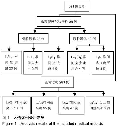
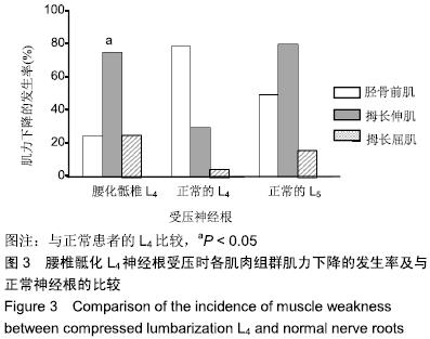
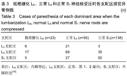
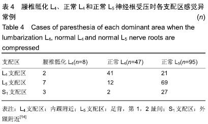
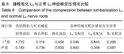
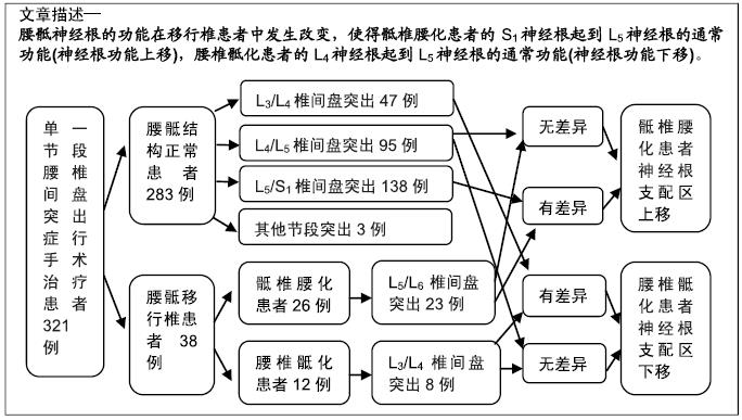
方法:研究方案的实施符合滨州医学院附属医院对研究的相关伦理要求,参与试验的患病个体及其家属对试验过程完全知情同意。回顾分析321例单一节段腰椎间盘突出症行手术治疗患者的病历资料。其中38例(11.8%)存在腰骶移行椎,包括骶椎腰化26例、腰椎骶化12例。26例骶椎腰化患者中,23例为L5/S1(L6)椎间盘突出,压迫S1(L6)神经根。12例腰椎骶化患者中,8例为L3/4椎间盘突出,压迫L4神经根。在283例正常结构的患者中,138例患者L5/S1椎间盘突出压迫S1神经根,95例患者L4/L5椎间盘突出压迫L5神经根,47例患者L3/L4椎间盘突出压迫L4神经根。比较术前骶椎腰化患者S1神经根受压的症状、腰椎骶化患者L4神经根受压的症状与正常腰骶椎患者L4、L5或S1神经根受压的症状。

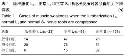
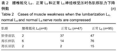
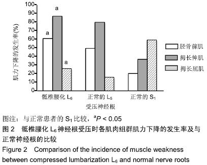
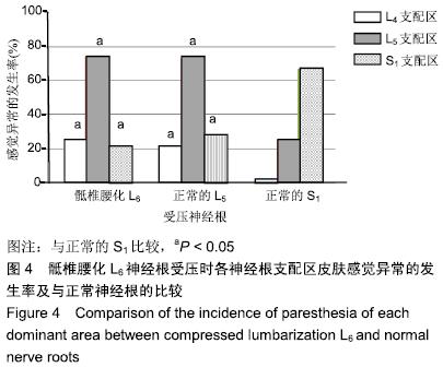
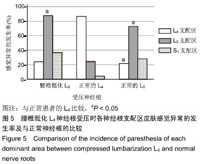
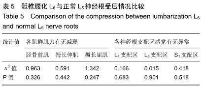
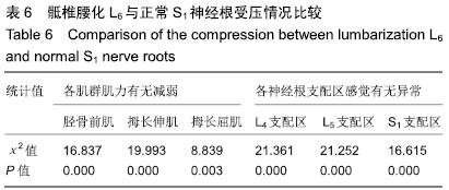
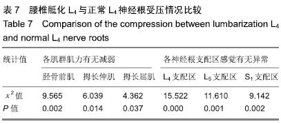
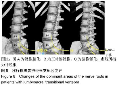
结果与结论:①S1神经根受压所致运动功能减退的分布在骶椎腰化患者组和正常组之间差异有显著性意义  (P < 0.05);②L4神经根受压所致运动功能减退的分布在腰椎骶化患者组和正常组之间差异有显著性意义(P < 0.05);③骶椎腰化患者S1神经根受压所致的运动功能减退与正常状态下L5神经根受压所致的运动功能减退相似;而腰椎骶化患者L4神经根受压所致的运动功能减退与正常状态下L5神经根受压所致的运动功能减退相似;皮肤感觉异常的分析也显示了相似的结果;④结果说明,腰骶神经根的功能在移行椎患者中发生改变,使得骶椎腰化患者的S1神经根起到L5神经根的通常功能(神经根功能上移),腰椎骶化患者的L4神经根起到L5神经根的通常功能(神经根功能下移)。

ORCID: 0000-0001-5041-2060(王欣)

中国组织工程研究杂志出版内容重点:组织构建;骨细胞;软骨细胞;细胞培养;成纤维细胞;血管内皮细胞;骨质疏松组织工程


关键词: 腰骶移行椎, 腰骶神经根, 支配区变化, 腰椎间盘突出

Abstract:

BACKGROUND: Lumbosacral transitional vertebra is a commonly seen congenital spinal deformity, and the changes in the muscle innervation pattern and the sensory dermatomes of the lumbosacral nerve roots have been reported, but the changes and its guidance significance for the surgeries of lumbar disc herniation have not been clarified systematically.

OBJECTIVE: To explore the possibility of changes in the muscle innervation pattern and the sensory dermatomes of the lumbosacral nerve roots when there is a lumbosacral transitional vertebra.

METHODS: The study was in accordance with the ethical requirements of Affiliated Hospital of Binzhou Medical University, and the subjects and their families signed the informed consents. The medical records of 321 patients with single segment lumbar disc herniation who underwent surgical treatment were analyzed retrospectively. Lumbosacral transitional vertebrae were present in 38 of 321 patients (11.8%). There were 26 cases of sacral lumbarization and 12 cases of lumbar sacralization. Among these 26 patients with sacral lumbarization, 23 had herniated discs at L5/S1 (L6) compressing the S1 (L6) nerve root. Of the 12 patients with lumbar sacralization, 8 had herniated discs at L3/4 compressing the L4 nerve root. In the 283 normally configured patients, 138 had herniated discs at L5/S1 compressing the S1 nerve root, 95 had herniated discs at L4/L5 compressing the L5 nerve root, and 47 had herniated discs at L3/L4 compressing the L4 nerve root. The preoperative symptoms of S1 nerve root compression in the patients with sacral lumbarization and of L4 nerve root compression in the patients with lumbar sacralization were compared with those of L4, L5 or S1 nerve root compression in the patients with normal configuration.

RESULTS AND CONCLUSION: (1) The distribution of motor function depression caused by S1 nerve root compression was significantly different between sacral lumbarization patients group and normal group (P < 0.05). (2) The distribution of motor function depression caused by L4 nerve root compression was also significantly different between lumbar sacralization patients group and normal group (P < 0.05). (3) The motor function depression caused by S1 nerve root compression in sacral lumbarization patients was similar to that of the L5 nerve root compression in the normal configuration, while the motor function depression caused by L4nerve root compression in lumbar sacralization patients was similar to that of the Lnerve root compression in the normal configuration. The analysis of the sensory dermatomes also showed similar results. (4) Our results suggest that the function of lumbosacral nerve roots changes in patients with transitional vertebrae. The S1 nerve roots in patients with sacral lumbarization tend to serve the usual function of L5 nerve roots (nerve roots move up), and the L4 nerve roots in patients with lumbar sacralization tend to serve the usual function of Lnerve roots (nerve roots move down).

Key words: lumbosacral transitional vertebrae, lumbosacral nerve roots, distribution variation, lumbar disc herniation

中图分类号: