中国组织工程研究 ›› 2020, Vol. 24 ›› Issue (3): 372-377.doi: 10.3969/j.issn.2095-4344.2411

• 骨科植入物 orthopedic implant • 上一篇    下一篇

跨骺钢板固定一定周期取出:继续观察一段时间后骺板的生长抑制情况

崔庆达1,王海革1,赵海军1,刘  伟2,毕郑钢2   

  1. 1青岛市胶州中心医院骨科,山东省青岛市  266300;2哈尔滨医科大学附属第一医院骨外一科,黑龙江省哈尔滨市  150000
  • 收稿日期:2019-03-18 修回日期:2019-03-28 接受日期:2019-04-30 出版日期:2020-01-28 发布日期:2019-12-25
  • 通讯作者: 毕郑钢,博士生导师,主任医师,哈尔滨医科大学附属第一医院骨外一科,黑龙江省哈尔滨市 150000
  • 作者简介:崔庆达,男,1982年生,山东省兰陵县人,汉族,2011年哈尔滨医科大学毕业,硕士,主治医师,主要从事关节周围骨折损伤方面的研究。
  • 基金资助:
    黑龙江省教育厅科学技术研究项目(12531370)

Removal of transepiphyseal plate after fixation for a certain period of time: growth inhibition of epiphyseal plate after a period of observation

Cui Qingda1, Wang Haige1, Zhao Haijun1, Liu Wei2, Bi Zhenggang2   

  1. 1Department of Joint Trauma, Jiaozhou Central Hospital, Qingdao 266300, Shandong Province, China; 2First Department of Orthopedics, the First Affiliated Hospital of Harbin Medical University, Harbin 150000, Heilongjiang Province, China
  • Received:2019-03-18 Revised:2019-03-28 Accepted:2019-04-30 Online:2020-01-28 Published:2019-12-25
  • Contact: Bi Zhenggang, Doctoral supervisor, Chief physician, First Department of Orthopedics, the First Affiliated Hospital of Harbin Medical University, Harbin 150000, Heilongjiang Province, China
  • About author:Cui Qingda, Master, Attending physician, Department of Joint Trauma, Jiaozhou Central Hospital, Qingdao 266300, Shandong Province, China
  • Supported by:
    the Science and Technology Research Project of Heilongjiang Provincial Education Department, No. 12531370

摘要:

文题释义:
骨骺损伤:骺板软骨细胞共分为4层,骨骺损伤后骺软骨的结构发生了一系列修复重建性改变,其中骺板软骨增生层和肥大细胞层的细胞增生、分化、代谢活跃。由于肥大细胞层细胞增大,基质稀少,形成了骺板中最薄弱部位,故受到压力时变化最为显著。
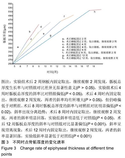
骺板厚度变化率:为随着时间延长,骺板厚度逐渐增加,单位时间内骺板厚度增长的比率。主要是指不同时间节点,骺板厚度生长变化情况。用于观察跨骺板钢板内固定一段时间,取出内固定物后骺板厚度的生长变化情况。

背景:临床上儿童干骺端、骺板周围骨折比较常见,跨骺板钢板内固定对稳定骨折有较为重要的作用。但内固定一定周期后取出内固定物,继续观察一定时间骺板的发育恢复情况鲜有报道。

目的:设计骺板周围骨折动物模型,分析跨骺板置入锁定钢板一定周期取出钢板,继续观察一段时间后骺板的生长抑制情况。

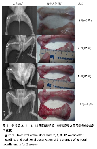
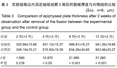
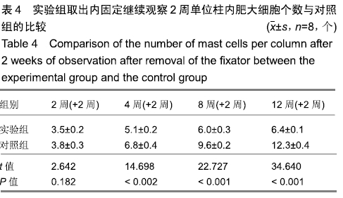
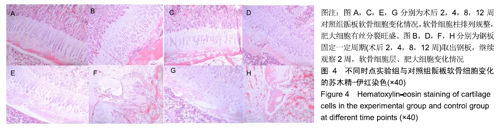
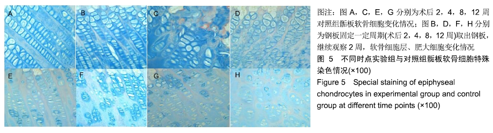
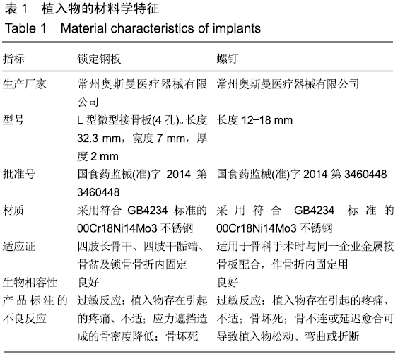
方法:建立32只幼兔右股骨远端骺板上方5 mm骨折模型,随机分4组,每组8只。应用相同型号钢板和螺钉,跨骺板周围骨折线行钢板置入内固定。术后2,4,8,12周取出内固定物,继续观察2周处死幼兔。取出股骨标本,测量股骨长度;作病理切片,测量骺板厚度及肥大细胞计数;观察形态学中肥大细胞及骺板厚度变化情况。将骨折模型作为实验组,以左股骨远端骺板作为对照。

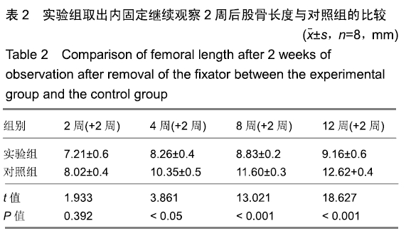
结果与结论:①实验组在内固定2周后,取出钢板继续观察2周,股骨长度、骺板厚度、肥大细胞计数等与对照组对比,差异无显著性意义;②实验组内固定4,8,12周,取出钢板继续观察2周,与对照组相比,实验组股骨长度、骺板厚度、肥大细胞计数等指标未能完全恢复至正常,差异有显著性意义(P < 0.05);③结果表明,在内固定物不伤及骺板的前提下,跨骺板钢板内固定初期(≤2周)取出钢板继续观察2周,适当压力作用对骺板生长发育未产生显著影响;④但持续过久压力限制时(≥4周),内固定对骺板压力限制时间过长,虽然也及时取出内固定,但肢体长度、骺板厚度、肥大细胞计数等指标均未能完全恢复正常,可导致骺板生长部分或者完全阻滞,引起肢体畸形及骺板发育停滞。

ORCID: 0000-0003-2448-9683(崔庆达)

中国组织工程研究杂志出版内容重点:人工关节;骨植入物;脊柱骨折;内固定;数字化骨科;组织工程

关键词: 骺板周围骨折, 跨骺板, 钢板, 内固定, 股骨长度, 肥大细胞, 动物实验

Abstract:

BACKGROUND: Clinically, children with metaphyseal and periepiphyseal fractures are more common. Internal fixation of the tarsal plate has a more important role in stabilizing the fracture. However, after a certain period of internal fixation, the fixator was taken out. After a certain period of observation, the recovery of the epiphyseal plate was rarely reported.

OBJECTIVE: To design an animal model of epiphyseal plate fracture and observe the growth and inhibition of epiphyseal plate after removal of the transepiphyseal steel plate implanted with locking plate for a period of time.

METHODS: The 5 mm fracture models of the distal epiphyseal plate of the right femur in 32 young rabbits were established. They were randomly divided into four groups with eight rats in each group. The same type of steel plate and screw was used. Internal fixation of transepiphyseal plate was conducted at periplate fracture line. The internal fixator was removed 2, 4, 8, and 12 weeks after operation. The rabbits were sacrificed after 2 weeks of observation. The femoral specimens were obtained, and the femoral length was measured. The thickness of epiphyseal plate and the number of mast cells were measured by pathological section. Morphological changes of mast cells and epiphyseal plate thickness were observed. The fracture model was used as the experimental group and the distal epiphyseal plate of the left femur was used as the control group.

RESULTS AND CONCLUSION: (1) After 2 weeks of internal fixation, the steel plate was removed in the experimental group and the observation was continued for 2 weeks. There was no significant difference in femoral length, epiphyseal thickness and mast cell count between the experimental group and the control group. (2) In the experimental group, the internal fixator was removed at 4, 8 and 12 weeks and the plate was observed for 2 weeks. Compared with the control group, the femoral length, epiphyseal thickness and mast cell count in the experimental group were not completely restored to normal, and the difference was significant (P < 0.05). (3) On the premise that the internal fixator did not injure the epiphyseal plate, the transepiphyseal plate was taken out at the initial stage of internal fixation (≤ 2 weeks), and the plate was observed for 2 additional weeks. The growth and development of the epiphyseal plate were not significantly affected by appropriate pressure. (4) If the pressure limitation lasts for too long (≥ 4 weeks), the time of internal fixation for epiphyseal plate pressure limitation is too long. Although the plate is removed in time, the indexes such as limb length, epiphyseal plate thickness and mast cell count cannot be completely restored to normal, which can lead to partial or complete blockade of epiphyseal plate growth, resulting in limb deformity and stagnation of epiphyseal plate development.

Key words: periepiphyseal fracture, transepiphyseal plate, plate, internal fixation, femoral length, mast cell, animal experiment

中图分类号: