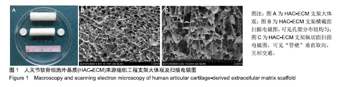
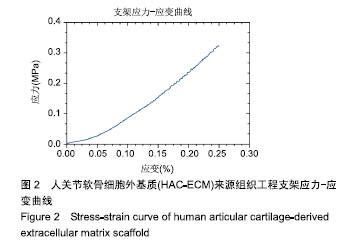
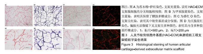
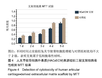
| [1]Johnstone B,Alini M,Cucchiarini M,et al.Tissue engineering for articular cartilage repair-the state of the art. Eur Cells Mater. 2013;25:248-267. [2]郭全义,肖统光,张一民,等.细胞外基质来源支架在软骨组织工程中的应用[J].中国组织工程研究, 2016,20(38):5737-5744.[3]Morille M, Toupet K, Montero-Menei CN, et al. PLGA-based microcarriers induce mesenchymal stem cell chondrogenesis and stimulate cartilage repair in osteoarthritis. Biomaterials. 2016;88:60-69.[4]Thiem A, Bagheri M, Grosse-Siestrup C, et al. Gelatin-poly(lactic-co-glycolic acid) scaffolds with oriented pore channel architecture - From in vitro to in vivo testing. Mater Sci Eng C Mater Biol Appl. 2016;62:585-595. [5]Xie X, Wang Y, Zhao C, et al. Comparative evaluation of MSCs from bone marrow and adipose tissue seeded in PRP-derived scaffold for cartilage regeneration. Biomaterials. 2012;33(29):7008-7018.[6]Almeida HV, Eswaramoorthy R, Cunniffe GM, et al. Fibrin hydrogels functionalized with cartilage extracellular matrix and incorporating freshly isolated stromal cells as an injectable for cartilage regeneration. Acta Biomater. 2016;36:55-62. [7]Sridhar BV, Dailing EA, Brock JL, et al. A Biosynthetic Scaffold that Facilitates Chondrocyte-Mediated Degradation and Promotes Articular Cartilage Extracellular Matrix Deposition. Regen Eng Transl Med. 2015;1(1-4):11-21. [8]Levingstone TJ, Ramesh A, Brady RT, et al. Cell- free multi-layered collagen-based scaffolds demonstrate layer specific regeneration of functional osteochondral tissue in caprine joints. Biomaterials. 2016;87:69-81.[9]Uematsu K, Hattori K, Ishimoto Y, et al. Cartilage regeneration using mesenchymal stem cells and a three-dimensional poly-lactic- glycolic acid (PLGA) scaffold. Biomaterials. 2005; 26(20):4273-4279.[10]Fan H, Hu Y, Zhang C, et al. Cartilage regeneration using mesenchymal stem cells and a PLGA-gelatin/ chondroitin/ hyaluronate hybrid scaffold. Biomaterials. 2006;27(26): 4573-4580.[11]Wang J, Yang Q, Cheng N, et al. Collagen/silk fibroin composite scaffold incorporated with PLGA microsphere for cartilage repair. Mater Sci Eng C Mater Biol Appl. 2016;61: 705-711. [12]Renth AN, Detamore MS.Leveraging "raw materials" as building blocks and bioactive signals in regenerative medicine.Tissue Eng Part B Rev. 2012;18(5):341-362. [13]Yang Q, Peng J, Guo Q, et al. A cartilage ECM-derived 3-D porous acellular matrix scaffold for in vivo cartilage tissue engineering with PKH26-labeled chondrogenic bone marrow-derived mesenchymal stem cells. Biomaterials. 2008;29(15):2378-2387. [14]Yang Q, Peng J, Lu SB, et al. Evaluation of an extracellular matrix-derived acellular biphasic scaffold/cell construct in the repair of a large articular high-load-bearing osteochondral defect in a canine model. Chin Med J (Engl). 2011;124(23): 3930-3938. [15]Zheng XF, Lu SB, Zhang WG, et al. Mesenchymal stem cells on a decellularized cartilage matrix for cartilage tissue engineering.Biotechnol Bioprocess Eng.2011;16(3):593-602.[16]肖统光,郝春香,荆晓光,等. 关节软骨细胞外基质/人脐带Wharton 胶复合多孔支架的制备及评估[J].中国组织工程研究,2017, 21(22):3470-3475. [17]Rezwan K, Chen QZ, Blaker JJ, et al. Biodegradable and bioactive porous polymer/inorganic composite scaffolds for bone tissue engineering. Biomaterials. 2006;27(18): 3413-3431. [18]Bhardwaj G, Webster TJ. Enhanced chondrocyte culture and growth on biologically inspired nanofibrous cell culture dishes. Int J Nanomedicine. 2016;11:479-483.[19]Sun Y, Yan L, Chen S, et al.Functionality of decellularized matrix in cartilage regeneration: A comparison of tissue versus cell sources.Acta Biomater. 2018;74:56-73.[20]Bradham DM, Passaniti A, Horton WE Jr.Mesenchymal cell chondrogenesis is stimulated by basement membrane matrix and inhibited by age-associated factors.Matrix Biol.1995; 14(7):561-571. [21]Gattazzo F, Urciuolo A, Bonaldo P.Extracellular matrix: a dynamic microenvironment for stem cell niche.Biochim Biophys Acta. 2014;1840(8):2506-2519. [22]Lynch K, Pei M.Age associated communication between cells and matrix: a potential impact on stem cell-based tissue regeneration strategies.Organogenesis. 2014;10(3):289-298. [23]Schneider C, Lehmann J, van Osch GJ, et al.Systematic Comparison of Protocols for the Preparation of Human Articular Cartilage for Use as Scaffold Material in Cartilage Tissue Engineering.Tissue Eng Part C Methods. 2016;22(12): 1095-1107.[24]Schneider KH, Enayati M, Grasl C, et al.Acellular vascular matrix grafts from human placenta chorion: Impact of ECM preservation on graft characteristics, protein composition and in vivo performance.Biomaterials. 2018;177:14-26. [25]Sivandzade F, Mashayekhan S. Design and fabrication of injectable microcarriers composed of acellular cartilage matrix and chitosan.J Biomater Sci Polym Ed. 2018;29(6):683-700. |
.jpg)

.jpg)
.jpg)