中国组织工程研究 ›› 2019, Vol. 23 ›› Issue (28): 4558-4563.doi: 10.3969/j.issn.2095-4344.1473
• 骨与关节图像与影像 bone and joint imaging • 上一篇 下一篇
4-6岁儿童下颈椎关节突关节的三维数字化形态特征
和雨洁1,张少杰1,李志军1,2,李筱贺1,王海燕1,2,王 星1,2,许阳阳1,高明杰1,李 琨1,戴丽娜1
- 内蒙古医科大学基础医学院,1人体解剖学教研室,2数字医学中心,内蒙古自治区呼和浩特市 010059
Three-dimensional digital morphological characteristics of the facet joint of the lower cervical spine in children aged 4-6 years
He Yujie1, Zhang Shaojie1, Li Zhijun1, 2, Li Xiaohe1, Wang Haiyan1, 2, Wang Xing1, 2, Xu Yangyang1, Gao Mingjie1, Li Kun1, Dai Lina1
- 1Department of Human Anatomy, 2Digital Medical Center, Basic Medical College, Inner Mongolia Medical University, Hohhot 010059, Inner Mongolia Autonomous Region, China
摘要:
文章快速阅读:
.jpg) 文题释义:
文题释义:儿童下颈椎:颈椎位于脊柱颈段,共7块,不仅支撑头的质量,还有很大的活动范围。C3-C7,因其解剖结构和功能与C1和C2有明显不同,故临床上习惯称为下颈椎。儿童颈部与成人颈部在解剖学、形态学方面存在较大差异,儿童颈部骨骼较纤细,韧带较松弛,关节面趋于水平,这些差异将导致儿童颈部相对成人更易受到损伤。
颈椎关节突关节的应用解剖:相邻椎骨的上、下关节突构成关节突关节,属于滑膜关节。颈椎关节突关节囊富含感受器,可感受生理刺激的强度,而且过度牵拉可产生疼痛感。关节突关节的面积、形态与关节的稳定有密切关系,成人C3的关节突关节面形态呈圆形,C4、C5逐渐改变为横椭圆形,C7、T1呈长横形,这种改变可能与适应颈椎生理运动关联,其大小、坡度上、下相适应,随脊柱节段不同而变化,以利于脊柱运动。
摘要
背景:颈椎关节突关节又称椎间小关节,与全身各种大关节一样属于滑膜关节。近年来解剖学和生物力学研究表明,颈椎关节突关节损伤、退变是引起慢性颈痛的最常见原因之一,现有研究主要集中于青少年和成人。
目的:测量儿童下颈椎关节突关节的相关形态参数,探讨其发育规律和形态特征,为临床经关节螺钉固定提供解剖学参数。
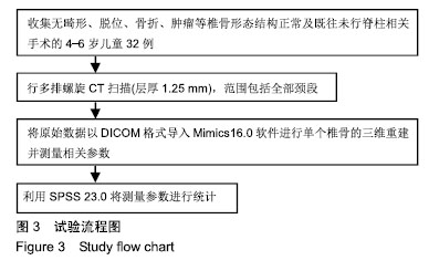
方法:选择4-6岁儿童32例,行下颈椎螺旋CT扫描后三维重建,要求无骨质破坏、肿瘤、畸形、骨折等,椎骨形态结构未发生改变,既往无脊柱相关手术,监护人对试验方案知情同意,且得到医院伦理委员会批准。进行C3-C7关节突关节相关形态测量及统计分析。
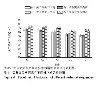
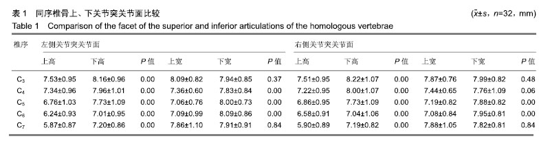
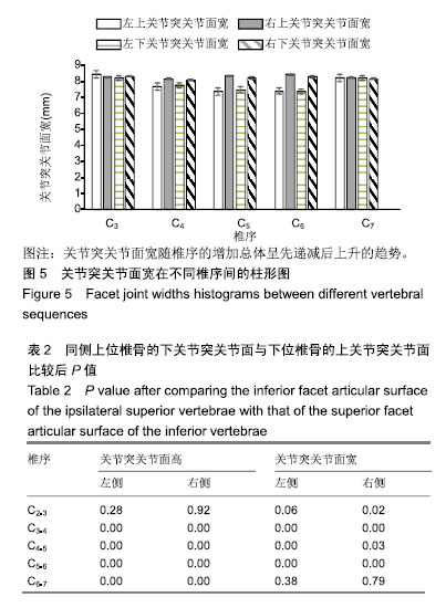
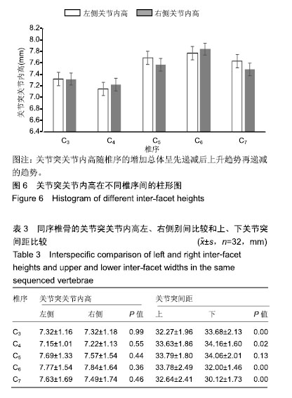
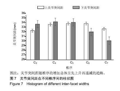
结果与结论:①儿童颈椎关节突关节面无论高度还是宽度,在相邻两椎骨之间总是下关节面较上关节面小,由此推断出关节面的大小总是上关节面大于下关节面,根据高宽比例得出4-6岁儿童下颈椎上关节突关节面从C3-C7皆接近椭圆形,下关节突关节面从C3-C7皆接近圆形,还未形成从圆形到椭圆形的过渡改变;②关节突关节内高变化趋势从C2-C7在C4节段出现最低点,总体呈先降后升趋势;③关节突间距先升后降,上关节突间距与下关节突间距除在C5节段之间差异无显著性意义且达最大值,在C3和C4节段上关节突间距均小于下关节突间距,在C6和C7节段上关节突间距明显大于下关节突间距,这可能与重力传递和颈椎活动有关。
ORCID: 0000-0002-1977-3180(和雨洁)
中图分类号:
.jpg)
.jpg) 文题释义:
文题释义: