中国组织工程研究 ›› 2019, Vol. 23 ›› Issue (27): 4344-4349.doi: 10.3969/j.issn.2095-4344.1383
• 精品专题 special topic • 上一篇 下一篇
雄激素干预成年雄性大脑中动脉阻断模型大鼠脑组织Bcl-2、Bax与Cyt-C的表达
龙艳芳1,2,王新蕾2,王明璞3,唐兴江2
- (1遂宁市中心医院康复科,四川省遂宁市 629000;2西南医科大学附属医院神经内科,四川省泸州市 646000;3遂宁市第一人民医院胸外科,四川省遂宁市 629000)
Effects of androgen on the expression of Bcl-2, Bax and Cyt-C in brain tissue of adult rat models of middle cerebral artery occlusion
Long Yanfang1, 2, Wang Xinlei2, Wang Mingpu3, Tang Xingjiang2
- (1Department of Rehabilitation Medicine, Suining Central Hospital, Suining 629000, Sichuan Province, China; 2Department of Neurology, the Affiliated Hospital of Southwest Medical University, Luzhou 646000, Sichuan Province, China; 3Department of Thoracic Surgery, Suining First People’s Hospital, Suining 629000, Sichuan Province, China)
摘要:
文章快速阅读:
.jpg) 文题释义:
脑缺血再灌注损伤:对于缺血性脑血管病而言,挽救缺血半暗带细胞的关键在于尽早缓解血管闭塞程度以恢复脑组织的血流供应,迄今临床上以溶栓为主要的应对手段。然而,脑缺血一定程度之后临床在使血流再通的同时出现难以避免的甚至更为严峻的脑细胞再灌注损伤,这种情况称之为脑缺血再灌注损伤。
细胞凋亡:指机体为维持内环境稳定,由基因控制的细胞自主的有序的死亡。与细胞坏死不同的是,细胞凋亡不是一件被动的过程,而是主动过程,它涉及一系列基因的激活、表达以及调控等的作用,它并不是病理条件下自体损伤的一种现象,而是为更好地适应生存环境而主动争取的一种死亡过程。它在生物体的进化、内环境的稳定以及多个系统的发育中起着重要的作用。
文题释义:
脑缺血再灌注损伤:对于缺血性脑血管病而言,挽救缺血半暗带细胞的关键在于尽早缓解血管闭塞程度以恢复脑组织的血流供应,迄今临床上以溶栓为主要的应对手段。然而,脑缺血一定程度之后临床在使血流再通的同时出现难以避免的甚至更为严峻的脑细胞再灌注损伤,这种情况称之为脑缺血再灌注损伤。
细胞凋亡:指机体为维持内环境稳定,由基因控制的细胞自主的有序的死亡。与细胞坏死不同的是,细胞凋亡不是一件被动的过程,而是主动过程,它涉及一系列基因的激活、表达以及调控等的作用,它并不是病理条件下自体损伤的一种现象,而是为更好地适应生存环境而主动争取的一种死亡过程。它在生物体的进化、内环境的稳定以及多个系统的发育中起着重要的作用。摘要
背景:研究发现,在脑梗死急性期无论男女均存在不同程度的性激素失衡,在男性则主要表现为睾酮下降、雌二醇升高,随着病情的恢复逐渐趋于正常。
目的:探讨雄激素对脑缺血再灌注损伤后脑组织Bcl-2、Bax与Cyt-C表达的影响。
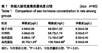
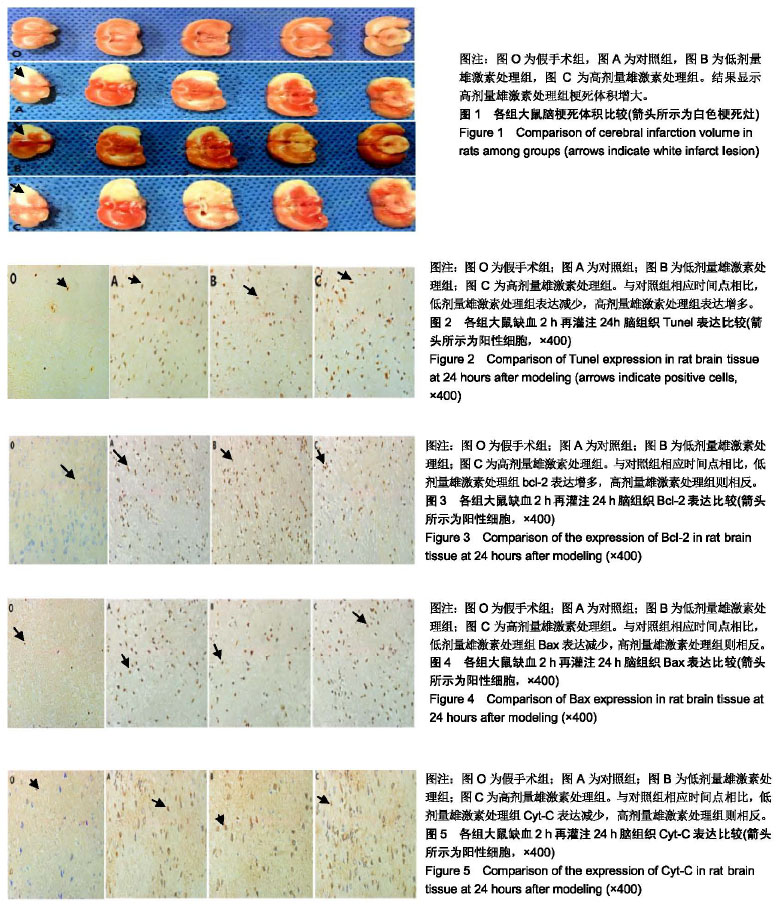
方法:120只健康成年雄性SD大鼠由西南医科大学动物实验中心提供,实验方案经西南医科大学动物实验伦理委员会批准(批准号为20170821026)。将120只大鼠随机分为假手术组、对照组及低、高剂量雄激素处理组(低剂量组、高剂量组)。应用改良Longa法制作大脑中动脉阻断模型大鼠,假手术组不进行插线操作。据脑缺血2 h后再灌注时间点的不同每组再随机分为5个亚组(6,12,24,48,72 h),每亚组5只。24 h时每组另有5只大鼠用于测量脑梗死体积。应用TUNEL法测原位凋亡细胞,免疫组织化学法检测Bcl-2、Bax与Cyt-C的表达情况。
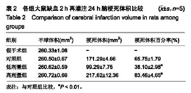
结果与结论:①假手术组未见梗死灶,原位凋亡细胞及Bcl-2、Bax与Cyt-C的表达较少,且无动态变化;与对照组相比,低剂量组脑梗死体积百分率、凋亡细胞及Bax与Cyt-C的表达减少,Bcl-2表达增多(均P < 0.05),高剂量组则相反(均P < 0.05);②除假手术组外,其他3组Bcl-2与Bax于脑缺血再灌注损伤后6 h表达逐渐增强,并于24 h达到高峰,随后逐渐下降;③结果说明,脑缺血再灌注损伤后,低剂量雄激素可能通过减少Bax与Cyt-C的表达、增强Bcl-2的表达,进而减少细胞凋亡,发挥神经保护作用,高剂量则作用相反。
中图分类号:


.jpg)
.jpg) 文题释义:
脑缺血再灌注损伤:对于缺血性脑血管病而言,挽救缺血半暗带细胞的关键在于尽早缓解血管闭塞程度以恢复脑组织的血流供应,迄今临床上以溶栓为主要的应对手段。然而,脑缺血一定程度之后临床在使血流再通的同时出现难以避免的甚至更为严峻的脑细胞再灌注损伤,这种情况称之为脑缺血再灌注损伤。
细胞凋亡:指机体为维持内环境稳定,由基因控制的细胞自主的有序的死亡。与细胞坏死不同的是,细胞凋亡不是一件被动的过程,而是主动过程,它涉及一系列基因的激活、表达以及调控等的作用,它并不是病理条件下自体损伤的一种现象,而是为更好地适应生存环境而主动争取的一种死亡过程。它在生物体的进化、内环境的稳定以及多个系统的发育中起着重要的作用。
文题释义:
脑缺血再灌注损伤:对于缺血性脑血管病而言,挽救缺血半暗带细胞的关键在于尽早缓解血管闭塞程度以恢复脑组织的血流供应,迄今临床上以溶栓为主要的应对手段。然而,脑缺血一定程度之后临床在使血流再通的同时出现难以避免的甚至更为严峻的脑细胞再灌注损伤,这种情况称之为脑缺血再灌注损伤。
细胞凋亡:指机体为维持内环境稳定,由基因控制的细胞自主的有序的死亡。与细胞坏死不同的是,细胞凋亡不是一件被动的过程,而是主动过程,它涉及一系列基因的激活、表达以及调控等的作用,它并不是病理条件下自体损伤的一种现象,而是为更好地适应生存环境而主动争取的一种死亡过程。它在生物体的进化、内环境的稳定以及多个系统的发育中起着重要的作用。