中国组织工程研究 ›› 2019, Vol. 23 ›› Issue (1): 30-34.doi: 10.3969/j.issn.2095-4344.1522

• 骨髓干细胞 bone marrow stem cells • 上一篇    下一篇

骨形态发生蛋白2基因转染骨髓间充质干细胞复合纤维蛋白胶促进前交叉韧带重建后的腱-骨愈合

朱乐全,卢剑华,冉俊岭,杨卫保,王 辉   

  1. 重庆市黔江中心医院·吉首大学附属黔江医院骨科,重庆市 409000
  • 修回日期:2018-09-05 出版日期:2019-01-08 发布日期:2018-11-28
  • 通讯作者: 卢剑华,硕士,重庆市黔江中心医院·吉首大学附属黔江医院骨科,重庆市 409000
  • 作者简介:朱乐全,男,汉族,1982年生,2009年重庆医科大学毕业,硕士,主要从事关节外科、创伤外科方面的研究。

Bone morphogenetic protein 2 transfected bone marrow mesenchymal stem cells and fibrin glue to promote tendon-bone healing after anterior cruciate ligament reconstruction

Zhu Lequan, Lu Jianhua, Ran Junling, Yang Weibao, Wang Hui   

  1. Department of Orthopedics, Qianjiang Central Hospital of Chongqing (Qianjiang Hospital of Jishou University), Chongqing 409000, China
  • Revised:2018-09-05 Online:2019-01-08 Published:2018-11-28
  • Contact: Lu Jianhua, Master, Department of Orthopedics, Qianjiang Central Hospital of Chongqing (Qianjiang Hospital of Jishou University), Chongqing 409000, China
  • About author:Zhu Lequan, Master, Department of Orthopedics, Qianjiang Central Hospital of Chongqing (Qianjiang Hospital of Jishou University), Chongqing 409000, China

摘要:

文章快速阅读:

文题释义:
腱-骨愈合:
指肌腱与骨道之间的生长愈合,以及相应的组织改变过程,其特征性结构为移行的纤维软骨带;腱-骨愈合方式大体上可分为直接愈合和间接愈合。
骨形态发生蛋白:是广泛存在于骨基质中的酸性多肽,具有明显的诱导成骨活性。实验中应用的骨形态发生蛋白2是研究最广泛且诱导成骨效果最明显的生长因子,在骨折、骨创伤、骨缺损等基础与临床试验中都显示了良好的效果,但是其单独应用存在半衰期短、降解过快的问题。


摘要
背景:
骨髓间充质干细胞与纤维蛋白胶联合应用可提高前交叉韧带重建后的早期腱-骨界面断裂强度与刚度,促进移植物与骨隧道早期愈合。
目的:探讨骨形态发生蛋白2基因转染骨髓间充质干细胞复合纤维蛋白胶对前交叉韧带重建术后腱-骨愈合的影响。
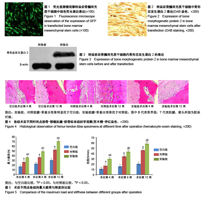
方法:将腺病毒载体Ad-GFP-骨形态发生蛋白2转染至骨髓间充质干细胞,48 h后检测GFP基因表达及骨形态发生蛋白2表达。将54只新西兰大白兔(购买于重庆市医学实验动物中心)随机分为空白组、实验组、对照组,每组18只,均建立前交韧带重建模型,实验组将基因转染48 h的骨髓间充质干细胞与纤维蛋白胶混合,注射至腱-骨界面;对照组将未转染的骨髓间充质干细胞与纤维蛋白胶混合,注射至腱-骨界面;空白组未注射任何材料。术后第4,8,12周分别获取股骨-移植肌腱-胫骨标本,进行组织学检测与生物力学检测。
结果与结论:①转染48 h后,荧光显微镜下可见细胞表达绿色荧光,转染效率为(86.1±6.3)%;SP免疫组织化学检测细胞胞浆内骨形态发生蛋白表达阳性;Western-blot检测可见骨形态发生蛋白2阳性表达条带;②苏木精-伊红染色显示,实验组、对照组腱-骨愈合效果明显优于空白组,实验组腱-骨愈合效果优于对照组;③生物力学检测显示,实验组、对照组的最大载荷、刚度均强于空白组(P < 0.05),实验组的最大载荷、刚度均强于对照组(P < 0.05);④结果表明,骨形态发生蛋白2进一步增强了骨髓间充质干细胞复合纤维蛋白胶促进前交叉韧带重建后的腱-骨愈合效果。

中国组织工程研究杂志出版内容重点:干细胞;骨髓干细胞;造血干细胞;脂肪干细胞;肿瘤干细胞;胚胎干细胞;脐带脐血干细胞;干细胞诱导;干细胞分化;组织工程
ORCID:
0000-0001-7055-4875(朱乐全)

关键词: 前交叉韧带重建, 骨形态发生蛋白2, 骨髓间充质干细胞, 纤维蛋白胶, 腱-骨愈合, 干细胞

Abstract:

BACKGROUND: The combination of bone marrow mesenchymal stem cells and fibrin glue can improve the early rupture strength and stiffness of the tendon-bone interface after anterior cruciate ligament reconstruction, and promote the early healing of graft and bone tunnel.
OBJECTIVE: To explore the effect of bone morphogenetic protein 2 transfected bone marrow mesenchymal stem cells combined with fibrin glue on the tendon-bone healing after the reconstruction of the anterior cruciate ligament.
METHODS: Adenovirus vector Ad-GFP-bone morphogenetic protein 2 was transfected into bone marrow mesenchymal stem cells, and the expressions of GFP gene and bone morphogenetic protein 2 were detected after 48 hours. Fifty-four New Zealand white rabbits purchased from the Medical Animal Center of Chongqing were randomly divided into blank group, experimental group and control group, with 18 rats in each group. The anterior cruciate ligament reconstruction model was established in each rat. In the experimental group, after 48 hours of transfection, the transfected bone marrow mesenchymal stem cells were mixed with fibrin glue and injected into the tendon-bone interface. In the control group, non-transfected bone marrow mesenchymal stem cells were mixed with fibrin glue and injected into the tendon-bone interface. In the blank group, no treatment was given. After 4, 8, 12 weeks, the femur-tendon-tibia specimens were taken for histological and biomechanical examinations.
RESULTS AND CONCLUSION: (1) After transfection of 48 hours, the cells expressed green fluorescence under the fluorescence microscope, and the transfection efficiency was (86.1±6.3)%. The expression of cytoplasmic bone morphogenetic protein was positive as shown by SP immunohistochemical test, and the positive band of bone morphogenetic protein 2 was detected by western blot. (2) Findings from the hematoxylin-eosin staining showed best better tendon-bone healing in the experimental and control group than the blank group, and the tendon-bone healing was further better in the experimental group than in the control group. (3) The biomechanical results showed that the maximum load and stiffness were ranked as follows: the experimental group > control group > blank group (P < 0.05). Therefore, bone morphogenetic protein 2-transfected bone marrow mesenchymal stem cells combined with fibrin glue can further enhance the tendon-bone healing after anterior cruciate ligament reconstruction. 

中国组织工程研究杂志出版内容重点:干细胞;骨髓干细胞;造血干细胞;脂肪干细胞;肿瘤干细胞;胚胎干细胞;脐带脐血干细胞;干细胞诱导;干细胞分化;组织工程

Key words: Anterior Cruciate Ligament, Bone Morphogenetic Proteins, Bone Marrow, Mesenchymal Stem Cells, Fibrin Tissue Adhesive, Tissue Engineering

中图分类号: