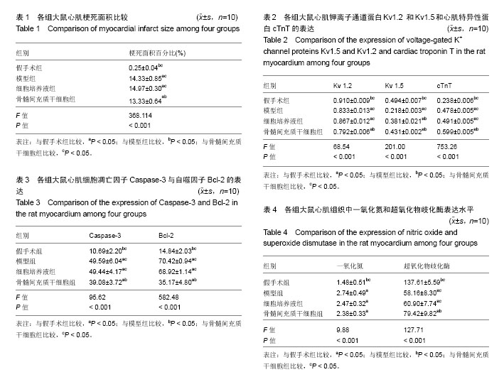
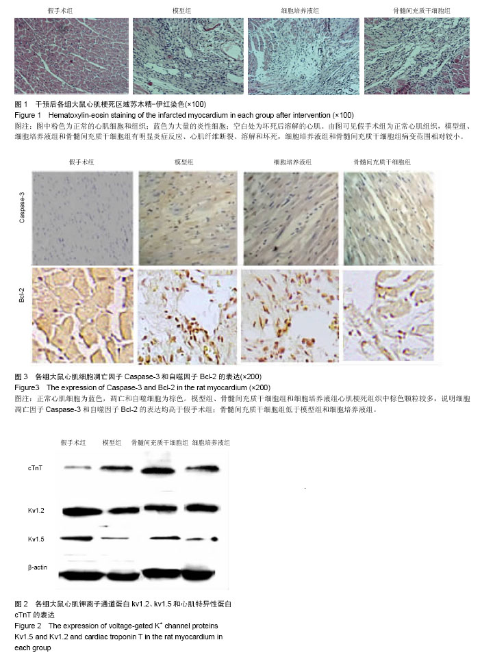
| [1] 吕光,雷建华,曹秀云.不同年龄组急性心肌梗死危险因素临床分析[J].中国实用医药,2008,3(28):91-92.[2] Brunskill SJ, Hyde CJ, Doree CJ, et al. Route of delivery and baseline left ventricular ejection fraction, key factors of bone-marrow-derived cell therapy for ischaemic heart disease. Eur J Heart Fail. 2009;11(9):887-896.[3] 罗涛,李玉明.心肌梗死后心室电重构的细胞核分子生物学机制[J].中国病理生理杂志,2008,24(11):2276-2281.[4] 杨琳,黄诒焯. 心肌细胞离子通道与心律失常[J].中国心脏起搏与心电生理杂志,2003,17(2):81-88.[5] Deng XL, Sun HY, Lau CP, et al. Properties of ion channels in rabbit mesenchymal stem cells from bone marrow. Biochem Biophys Res Commun. 2006;348(1):301-309.[6] Cai B, Wang G, Chen N, et al. Bone marrow mesenchymal stem cells protected post-infarcted myocardium against arrhythmias via reversing potassium channels remodelling. J Cell Mol Med. 2014;18(7):1407-1416.[7] Pfeffer MA, Pfeffer JM, Steinberg C, et al. Survival after an experimental myocardial infarction: beneficial effects of long-term therapy with captopril. Circulation. 1985;72(2): 406-412.[8] Gao L, Bledsoe G, Yin H, et al. Tissue kallikrein-modified mesenchymal stem cells provide enhanced protection against ischemic cardiac injury after myocardial infarction. Circ J. 2013;77(8):2134-2144.[9] Takagawa J, Zhang Y, Wong ML, et al. Myocardial infarct size measurement in the mouse chronic infarction model: comparison of area- and length-based approaches. J Appl Physiol (1985). 2007;102(6):2104-2111.[10] 杨国勋,刘唐威,钟国强,等.骨髓间充质干细胞移植对心肌梗死后心功能的影响[J]. 中华危重病急救医学, 2007, 19(7): 428-430.[11] 梁丽玲,杨庭树,李萍,等. 同种异体骨髓间充质干细胞移植治疗大鼠急性心肌梗死[J]. 中国组织工程研究,2014,18(37): 5983-5987.[12] Chen H, Xia R, Li Z, et al. Mesenchymal Stem Cells Combined with Hepatocyte Growth Factor Therapy for Attenuating Ischaemic Myocardial Fibrosis: Assessment using Multimodal Molecular Imaging. Sci Rep. 2016;6:33700. [13] Hua P, Liu LB, Liu JL, et al. Inhibition of apoptosis by knockdown of caspase-3 with siRNA in rat bone marrow mesenchymal stem cells. Exp Biol Med (Maywood). 2013; 238(9):991-998.[14] Li L, Guan Q, Dai S, et al. Integrin β1 Increases Stem Cell Survival and Cardiac Function after Myocardial Infarction. Front Pharmacol. 2017;8:135.[15] 李艳菊,刘洋,郭坤元.骨髓间充质干细胞对衰老心脏血管内皮生长因子表达的影响[J].中国组织工程研究,2012,16(41): 7709-7712.[16] Kheloufi M, Rautou PE, Boulanger CM. Autophagy in the cardiovascular system. Med Sci (Paris). 2017;33(3):283-289.[17] Lee SI, Kim BG, Hwang DH, et al. Overexpression of Bcl-XL in human neural stem cells promotes graft survival and functional recovery following transplantation in spinal cord injury. J Neurosci Res. 2009;87(14):3186-3197.[18] De Meyer GR, Martinet W. Autophagy in the cardiovascular system. Biochim Biophys Acta. 2009;1793(9):1485-1495.[19] Zhong Z, Hu J, Sun Y, et al. Impact of mesenchymal stem cells transplantation on myocardial myocardin-related transcription factor-A and bcl-2 expression in rats with experimental myocardial infarction. Zhonghua Xin Xue Guan Bing Za Zhi. 2015;43(6):531-536.[20] 吕延杰,蔡本志,艾静,等.骨髓间充质干细胞对心肌细胞K+电流的影响[J].中国药理通讯,2013,30(3):46.[21] Deng XL, Lau CP, Lai K, et al. Cell cycle-dependent expression of potassium channels and cell proliferation in rat mesenchymal stem cells from bone marrow. Cell Prolif. 2007;40(5):656-670.[22] Leanza L, Zoratti M, Gulbins E, et al. Induction of apoptosis in macrophages via Kv1.3 and Kv1.5 potassium channels. Curr Med Chem. 2012;19(31):5394-5404. |
.jpg)


.jpg)