[1] KEENEY M, CHUNG MT, ZIELINS ER, et al. Scaffold-mediated BMP-2 minicircle DNA delivery accelerated bone repair in a mouse critical-size calvarial defect model. J Biomed Mater Res A. 2016;104(8):2099-2107.
[2] 兰伟伟,陈维毅,黄棣.骨软骨组织工程研究进展[J].生物医学工程学杂志,2019,36(3):504-510.
[3] ATASOY-ZEYBEK A, KOSE GT. Gene Therapy Strategies in Bone Tissue Engineering and Current Clinical Applications. Adv Exp Med Biol. 2018; 1119: 85-101.
[4] 张爽,刘石,汪宇峰,等.间充质干细胞修复骨关节炎软骨损伤的临床应用意义[J].中国组织工程研究,2018,22(33):5379-5385.
[5] DAI R, WANG Z, SAMANIPOUR R, et al. Adipose-Derived Stem Cells for Tissue Engineering and Regenerative Medicine Applications. Stem Cells Int. 2016;2016:6737345.
[6] HAO W, LIU H, ZHOU L, et al. MiR-145 regulates osteogenic differentiation of human adipose-derived mesenchymal stem cells through targeting FoxO1. Exp Biol Med (Maywood). 2018;243(4):386-393.
[7] ZHANG Z, MA Y, GUO S, et al. Low-intensity pulsed ultrasound stimulation facilitates in vitro osteogenic differentiation of human adipose-derived stem cells via up-regulation of heat shock protein (HSP)70, HSP90, and bone morphogenetic protein (BMP) signaling pathway. Biosci Rep. 2018; 38(3):BSR20180087.
[8] UDDIN A, CHAKRABORTY S. Role of miRNAs in lung cancer. J Cell Physiol. 2018 Apr 20. doi: 10.1002/jcp.26607.
[9] LOPEZ-BERTONI H, KOZIELSKI KL, RUI Y, et al. Bioreducible Polymeric Nanoparticles Containing Multiplexed Cancer Stem Cell Regulating miRNAs Inhibit Glioblastoma Growth and Prolong Survival. Nano Lett. 2018;18(7): 4086-4094.
[10] HUANG S, WANG S, BIAN C, et al. Upregulation of miR-22 promotes osteogenic differentiation and inhibits adipogenic differentiation of human adipose tissue-derived mesenchymal stem cells by repressing HDAC6 protein expression. Stem Cells Dev. 2012;21(13):2531-2540.
[11] QURESHI AT, MONROE WT, DASA V, et al. miR-148b-nanoparticle conjugates for light mediated osteogenesis of human adipose stromal/stem cells. Biomaterials. 2013;34(31):7799-7810.
[12] WANG Z, XIE Q, YU Z, et al. A regulatory loop containing miR-26a, GSK3β and C/EBPα regulates the osteogenesis of human adipose-derived mesenchymal stem cells. Sci Rep. 2015;5:15280.
[13] ZHANG WB, ZHONG WJ, WANG L. A signal-amplification circuit between miR-218 and Wnt/β-catenin signal promotes human adipose tissue-derived stem cells osteogenic differentiation. Bone. 2014;58:59-66.
[14] HAO X, XIA L, QU R, et al. Association between miR-146a rs2910164 polymorphism and specific cancer susceptibility: an updated meta-analysis. Fam Cancer. 2018;17(3):459-468.
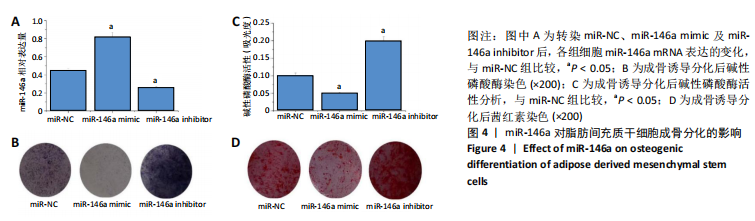
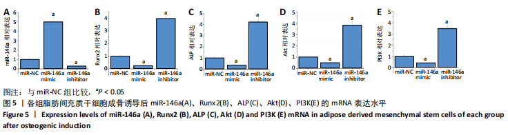
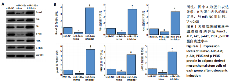
[15] 匡威,谭家莉,张红梅,等.miR-146a调控骨髓间充质干细胞成骨分化的机制研究[J].生物医学工程与临床,2011,15(5):413-416.
[16] MA M, WANG X, CHEN X, et al. MicroRNA-432 targeting E2F3 and P55PIK inhibits myogenesis through PI3K/AKT/mTOR signaling pathway. RNA Biol. 2017;14(3):347-360.
[17] LUO G, XU B, HUANG Y. Icariside II promotes the osteogenic differentiation of canine bone marrow mesenchymal stem cells via the PI3K/AKT/mTOR/S6K1 signaling pathways. Am J Transl Res. 2017;9(5):2077-2087.
[18] 李洋,陈前昭,邵英,等.骨形态蛋白9诱导干细胞骨向分化与环氧酶-2及PI3K/Akt的关系研究[J].中国药理学通报,2017,33(7): 908-915.
[19] 支力强,杨一欣,许毛,等.SIRT1通过PI3K/AKT通路促进成骨细胞分化的相关机制研究[J].实用骨科杂志,2018,24(6):523-526.
[20] 王雪鹏,李茂强,边振宇,等. PI3K/Akt信号通路在骨髓间充质干细胞增殖及成骨分化调控中的作用[J].中华骨质疏松和骨矿盐疾病杂志,2014,7(3):250-257.
[21] LI J, HU C, HAN L, et al. MiR-154-5p regulates osteogenic differentiation of adipose-derived mesenchymal stem cells under tensile stress through the Wnt/PCP pathway by targeting Wnt11. Bone. 2015;78: 130-141.
[22] LANGER R, VACANTI JP. Tissue engineering. Science. 1993;260(5110): 920-926.
[23] BAJEK A, GURTOWSKA N, OLKOWSKA J, et al. Adipose-Derived Stem Cells as a Tool in Cell-Based Therapies. Arch Immunol Ther Exp (Warsz). 2016;64(6):443-454.
[24] CHEN QJ, CHEN L, WU SK, et al. rhPDGF-BB combined with ADSCs in the treatment of Achilles tendinitis via miR-363/PI3 K/Akt pathway. Mol Cell Biochem. 2018;438(1-2):175-182.
[25] KRZESNIAK NE, SARNOWSKA A, FIGIEL-DABROWSKA A, et al. Secondary release of the peripheral nerve with autologous fat derivates benefits for functional and sensory recovery. Neural Regen Res. 2021;16(5):856-864.
[26] 刘润恒,刘于冬,陈卓凡.微小RNA在骨分化过程中的作用机制[J].国际口腔医学杂志,2017,44(1):108-113.
[27] QIAO L, LIU D, LI CG, et al. MiR-203 is essential for the shift from osteogenic differentiation to adipogenic differentiation of mesenchymal stem cells in postmenopausal osteoporosis. Eur Rev Med Pharmacol Sci. 2018;22(18):5804-5814.
[28] KIM YJ, BAE SW, YU SS, et al. miR-196a regulates proliferation and osteogenic differentiation in mesenchymal stem cells derived from human adipose tissue. J Bone Miner Res. 2009;24(5):816-825.
[29] LACCI KM, DARDIK A. Platelet-rich plasma: support for its use in wound healing. Yale J Biol Med. 2010;83(1):1-9.
[30] FKIH M’HAMED I, PRIVAT M, TRIMECHE M, et al. miR-10b, miR-26a, miR-146a And miR-153 Expression in Triple Negative Vs Non Triple Negative Breast Cancer: Potential Biomarkers. Pathol Oncol Res. 2017; 23(4):815-827.
[31] DONG Z, YU C, REZHIYA K, et al. Downregulation of miR-146a promotes tumorigenesis of cervical cancer stem cells via VEGF/CDC42/PAK1 signaling pathway. Artif Cells Nanomed Biotechnol. 2019;47(1): 3711-3719.
[32] HAN Z, CAO J, WANG Y, et al. Quercetin Suppresses Proliferation and Motility Through Modulating Hippo Pathway Via Upregulating Mir-146a-5p in Gastric Cancer. J Biomaterials Tissue Engineering. 2019;9:82-88.
[33] KIM DY, KIM EJ, JANG WG. Piperine induces osteoblast differentiation through AMPK-dependent Runx2 expression. Biochem Biophys Res Commun. 2018;495(1):1497-1502.
[34] NARAYANAN A, SRINAATH N, ROHINI M, et al. Regulation of Runx2 by MicroRNAs in osteoblast differentiation. Life Sci. 2019;232:116676.
[35] KHALID AB, SLAYDEN AV, KUMPATI J, et al. GATA4 Directly Regulates Runx2 Expression and Osteoblast Differentiation. JBMR Plus. 2018; 2(2):81-91.
[36] COSTA RLB, HAN HS, GRADISHAR WJ. Targeting the PI3K/AKT/mTOR pathway in triple-negative breast cancer: a review. Breast Cancer Res Treat. 2018;169(3):397-406.
[37] PORTA C, PAGLINO C, MOSCA A. Targeting PI3K/Akt/mTOR Signaling in Cancer. Front Oncol. 2014;4:64.
[38] ZHANG Y, YANG JH. Activation of the PI3K/Akt pathway by oxidative stress mediates high glucose-induced increase of adipogenic differentiation in primary rat osteoblasts. J Cell Biochem. 2013;114(11): 2595-2602. |