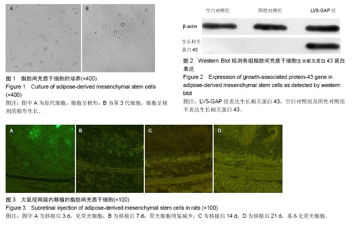
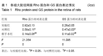
| [1] 王昕,张妍,张天资,等.病毒转染治疗视网膜色素变性研究进展[J].中国实用眼科杂志,2015,33(4):337-340.[2] Zhang Q.Retinitis Pigmentosa: Progress and Perspective. Asia Pac J Ophthalmol (Phila).2016;5(4):265-271. [3] 李光辉,曾芳,王晔恺,等.腺相关病毒载体在视网膜色素变性基因治疗中的应用研究进展[J].中华眼底病杂志,2014,30(6): 636-639.[4] Pensado A,Diaz-Corrales FJ,De la Cerda B,et al.Span poly-L-arginine nanoparticles are efficient non-viral vectors for PRPF31 gene delivery: An approach of gene therapy to treat retinitis pigmentosa.Nanomedicine.2016;12(8):2251-2260. [5] 杨欣悦,王晨光,苏冠方,等.生长相关蛋白43(GAP-43)与视神经视网膜损伤修复[J].中国实用眼科杂志,2015,33(8):853-856.[6] Zhu Q,Liu Z,Wang C,et al.Lentiviral-mediated growth-associated protein-43 modification of bone marrow mesenchymal stem cells improves traumatic optic neuropathy in rats.Mol Med Rep.2015;12(4):5691-5700. [7] He Y,Zhang Y,Liu X,et al.Recent advances of stem cell therapy for retinitis pigmentosa.Int J Mol Sci.2014;15(8): 14456-14474. [8] Li Y,Wu WH,Hsu CW,et al.Gene therapy in patient-specific stem cell lines and a preclinical model of retinitis pigmentosa with membrane frizzled-related protein defects.Mol Ther. 2014;22(9):1688-1697. [9] 郑祥榕,柳林,高朋芬,等.视网膜色素变性疾病中细胞替代治疗[J].中国实用眼科杂志,2013,31(8):953-956.[10] 刘玉平,刘涛,王明明,等.人髌下脂肪垫来源脂肪间充质干细胞的分离、培养及鉴定[J].中国组织工程研究,2015,19(41): 6566-6571.[11] Zhao Y,Zhang H.Update on the mechanisms of homing of adipose tissue-derived stem cells. Cytotherapy.2016; 18(7): 816-827. [12] 马洪斌,李运祥,王铭伦,等.腺病毒携带骨形态发生蛋白14基因转染脂肪干细胞修复损伤关节软骨?[J].中国组织工程研究,2015, 19(1):54-60.[13] Moradi SL,Eslami G,Goudarzi H,et al.Role of Helicobacter pylori on cancer of human adipose-derived mesenchymal stem cells and metastasis of tumor cells-an in vitro study. Tumour Biol.2016;37(3):3371-3378. [14] Petersen GF,Hilbert B,Trope G,et al.Efficient transduction of equine adipose-derived mesenchymal stem cells by VSV-G pseudotyped lentiviral vectors.Res Vet Sci.2014;97(3): 616-622.[15] Ghazi NG,Abboud EB,Nowilaty SR,et al.Treatment of retinitis pigmentosa due to MERTK mutations by ocular subretinal injection of adeno-associated virus gene vector: results of a phase I trial.Hum Genet.2016;135(3):327-343. [16] 基因治疗首次用于视网膜色素变性治疗[J].中国眼镜科技杂志, 2012,24(11):28-28.[17] Beltran WA,Cideciyan AV,Iwabe S,et al.Successful arrest of photoreceptor and vision loss expands the therapeutic window of retinal gene therapy to later stages of disease.Proc Natl Acad Sci U S A.2015;112(43):E5844-5853. [18] Mookherjee S,Hiriyanna S,Kaneshiro K,et al.Long-term rescue of cone photoreceptor degeneration in retinitis pigmentosa 2(RP2)-knockout mice by gene replacement therapy.Hum Mol Genet.2015;24(22):6446-6458. [19] 周朋义,彭广华.视网膜色素变性治疗的研究进展[J].眼科新进展, 2012,32(5):493-496. [20] Ng TK,Fortino VR,Pelaez D,et al.Progress of mesenchymal stem cell therapy for neural and retinal diseases.World J Stem Cells.2014;6(2):111-119.[21] Uy HS,Chan PS,Cruz FM.Stem cell therapy: a novel approach for vision restoration in retinitis pigmentosa.Med Hypothesis Discov Innov Ophthalmol.2013;2(2):52-55.[22] 李青兰.眼源性干细胞移植治疗视网膜疾病的研究进展[J].眼科新进展,2011,31(4):397-400. [23] 马倩倩,封志纯.骨髓间充质干细胞在视网膜病变治疗中的研究进展[J].中华实用儿科临床杂志,2015,30(18):1431-1433.[24] Wang Y,Yu X,Chen E,et al.Liver-derived human mesenchymal stem cells: a novel therapeutic source for liver diseases.Stem Cell Res Ther.2016;7(1):71.[25] Yamaoka K.Potential of bone regenerative therapy with mesenchymal stem cells in rheumatoid arthritis.Clin Calcium. 2016;26(5):758-762. [26] 郭宪立,刘跃,周利敏,等.脂肪间充质干细胞经肝动脉移植治疗晚期肝病[J].中国组织工程研究,2016,20(6):848-854.[27] 周金文,利焕廉,左炜,等.间充质干细胞对糖尿病病变模型大鼠血视网膜屏障的影响研究[J].亚太传统医药,2014,10(23):10-12.[28] Lye KL,Nordin N,Vidyadaran S,et al.Mesenchymal stem cells: From stem cells to sarcomas.Cell Biol Int.2016;40(6): 610-618. [29] Miyagi-Shiohira C,Kurima K,Kobayashi N,et al. Cryopreservation of Adipose-Derived Mesenchymal Stem Cells.Cell Med.2015;8(1-2):3-7.[30] 王勇,赵伟,冯健洲,等.神经生长因子修饰脂肪干细胞移植促进损伤脊髓的修复[J].中国组织工程研究,2015,19(14):2224-2229. [31] Abdul Halim NS,Fakiruddin KS,Ali SA,et al.A comparative study of non-viral gene delivery techniques to human adipose-derivedmesenchymal stem cell.Int J Mol Sci.2014; 15(9):15044-15060. [32] Yan X,Ehnert S,Culmes M,et al.5-azacytidine improves the osteogenic differentiation potential of aged human adipose-derivedmesenchymal stem cells by DNA demethylation. PLoS One.2014;9(6):e90846. [33] Moriyama H,Moriyama M,Sawaragi K,et al.Tightly regulated and homogeneous transgene expression in human adipose-derived mesenchymal stem cells by lentivirus with tet-off system.PLoS One.2013;8(6):e66274.[34] Kaneda M,Nagashima M,Nunome T,et al.Changes of phospho-growth-associated protein 43 (phospho-GAP43) in the zebrafish retina afteroptic nerve injury: a long-term observation.Neurosci Res.2008;61(3):281-288. [35] Williams RR,Venkatesh I,Pearse DD,et al.MASH1/Ascl1a leads to GAP43 expression and axon regeneration in the adult CNS.PLoS One.2015;10(3):e0118918. [36] 刘晓坤,罗钢,赵平.大鼠外伤性视神经病变后 GAP-43表达的变化[J].河北医药,2014,36(17):2568-2570. [37] 王瑞红,侯世科,吴志鸿,等.GAP-43与受损后视网膜神经节细胞存活和再生关系的研究进展[J].国际眼科纵览,2012, 36(3): 172-177. |
.jpg)
.jpg)