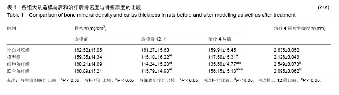
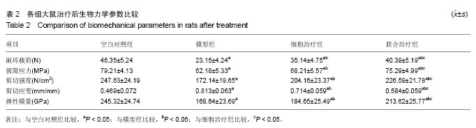
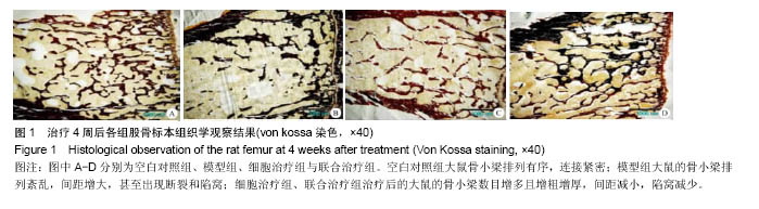
| [1]汪玉海,金丽娟,高俊,等.脂肪干细胞复合PLGA对骨质疏松骨折愈合后生物力学的影响[J]. 宁夏医科大学学报, 2013,35(3): 244-247.[2]Kim KI,Park S,Im GI.Osteogenic differentiation and angiogenesis with cocultured adipose-derived stromal cells and bone marrow stromal cells.Biomaterials.2014;35(17): 4792-4804.[3]Kim DW,Jeon BJ,Hwang NH,et al.Adipose-derived stem cells inhibit epidermal melanocytes through an interleukin-6-mediated mechanism.Plast Reconstr Surg. 2014;134(3):470-480. [4]王闯建.复合BMSCs包芯结构骨支架材料修复兔桡骨骨缺损的实验研究[D].西安:第四军医大学,2012.[5]Jeong HH,Piao S,Ha JN,et al.Combined therapeutic effect of udenafil and adipose-derived stem cell (ADSC)/brain-derived neurotrophic factor (BDNF)-membrane system in a rat model of cavernous nerve injury.Urology.2013;81(5):11087-11114.[6]韩玮,李煜明.密固达预防全髋关节置换术后骨质疏松临床研究[J].现代仪器与医疗,2015,21(4):119-120.[7]Wang W,Cao B,Cui L,et al.Adipose tissue engineering with human adipose tissue-derived adult stem cells and a novel porous scaffold.J Biomed Mater Res B Appl Biomater.2013; 101(1):68-75.[8]李鹏,黄成硕,高翔,等.大鼠骨髓间充质干细胞体外分离及对原发性骨质疏松性骨折的实验研究[J].国际检验医学杂志, 2015, 36(14):1984-1985.[9]周年,刘波,徐彭.氧化应激与骨质疏松症的研究进展[J].中国骨质疏松杂志,2014,20(12):1485-1489.[10]Karam JP,Muscari C,Sindji L,et al.Pharmacologically active microcarriers associated with thermosensitive hydrogel as a growth factor releasing biomimetic 3D scaffold for cardiac tissue-engineering.J Control Release.2014;192: 82-94. [11]Lee S,Lee IG,Kim AR,et al.Combined effects of brain-derived neurotrophic factor immobilized poly-lactic-co-glycolic acid membrane with human adipose-derived stem cells and basic fibroblast growth factor hydrogel on recovery of erectile dysfunction.Tissue Eng Part A.2014;20(17-18):2446-2454.[12]李春波,王红,陈增淦,等.兔脂肪干细胞(ADSCs)与聚羟基乙酸/壳聚糖(PLGA/CS)支架材料生物相容性研究[J].复旦学报(医学版),2014,41(5):610-616.[13]Kusuyama J,Bandow K,Shamoto M,et al.Low intensity pulsed ultrasound (LIPUS) influences the multilineage differentiation of mesenchymal stem and progenitor cell lines through ROCK-Cot/Tpl2-MEK-ERK signaling pathway.J Biol Chem. 2014;289(15):10330-10344.[14]林涛,陈竹,袁德超,等.壳聚糖水凝胶复合脂肪间充质干细胞修复兔关节软骨缺损[J].中华创伤杂志,2016,32(4):357-362.[15]Irmak G,Demirta? TT,Çetin Alt?ndal D,et al.Sustained release of 17β-estradiol stimulates osteogenic differentiation of adipose tissue-derived mesenchymal stem cells on chitosan-hydroxyapatite scaffolds.Cells Tissues Organs.2014; 199(1):37-50.[16]Zhang Q,Hubenak J,Iyyanki T,et al.Engineering vascularized soft tissue flaps in an animal model using human adipose-derived stem cells and VEGF+PLGA/PEG microspheres on a collagen-chitosan scaffold with a flow-through vascular pedicle.Biomaterials.2015;73:198-213.[17]于峥嵘,石旭东,李淳德,等.干细胞复合Ⅰ型胶原修饰的PLGA微球治疗骨质疏松性骨缺损[J].中华骨科杂志,2014,34(1):62-69.[18]Cruz AC,Caon T,Menin Á,et al.Adipose-Derived Stem Cells Decrease Bone Morphogenetic Protein Type 2-Induced Inflammation In Vivo.J Oral Maxillofac Surg. 2016;74(3): 505-514. [19]宋一民,蔺晨,李晓斌,等.体外PLGA三维支架上诱导小鼠骨髓干细胞向胰岛样细胞分化[J]. 国际生物医学工程杂志, 2014,37(5): 262-265.[20]刘锴,李春波,陈增淦,等.MRI示踪超顺磁性氧化铁标记兔脂肪干细胞治疗关节软骨缺损的研究[J].中国临床医学, 2015,22(5): 589-595.[21]Razavi S,Karbasi S,Morshed M,et al.Cell Attachment and Proliferation of Human Adipose-Derived Stem Cells on PLGA/Chitosan Electrospun Nano-Biocomposite.Cell J.2015;17(3):429-437.[22]Yin F,Cai J,Zen W,et al.Cartilage Regeneration of Adipose-Derived Stem Cells in the TGF-β1-Immobilized PLGA-Gelatin Scaffold.Stem Cell Rev.2015;11(3):453-459. [23]姜景进,石恒.脂肪源性干细胞(ADSCs)在美容整形外科应用的基本概念[J].医学与哲学, 2014,13(6):10-13.[24]钟晓敏,陈刚,张圣敏,等.重力矢量及趋化诱导对脂肪干细胞在PLGA电纺膜上早期增殖的影响[J].中国生物医学工程学报, 2014,33(4):508-512.[25]Chen DC,Chen LY,Ling QD,et al.Purification of human adipose-derived stem cells from fat tissues using PLGA/silk screen hybrid membranes.Biomaterials.2014; 35(14): 4278-4287.[26]韩晓谦,董志恒,于祥茹,等.载辛伐他汀PLGA/CPC复合骨髓基质干细胞构建组织工程骨的实验研究[J].上海口腔医学, 2014, 23(1):7-14.[27]许世兵,单乐天,金红婷,等.经血源子宫内膜干细胞复合3D打印PLGA支架体外培养的相容性研究[J].浙江中医药大学学报, 2015,39(12):854-858.[28]Vilquin JT, Rosset P.Mesenchymal stem cells in bone and cartilage repair: current status.Regen Med. 2006;1(4): 589-604.[29]Jung MS,Han BJ,Lee SE,et al.In vitro micro-mineralized tissue formation by the combinatory condition of adipose-derived stem cells, macroporous PLGA microspheres and a bioreactor.Macromol Res.2014;22(1):47-57.[30]王伟,阿里木江•阿不来提,艾尔肯•热合木吐拉,等.三维打印技术构建仿生人类指骨支架血管化的研究[J].中华实验外科杂志, 2014,31(7):1431-1433.[31]Akita D,Morokuma M,Saito Y,et al.Periodontal tissue regeneration by transplantation of rat adipose-derived stromal cells in combination with PLGA-based solid scaffolds.Biomed Res.2014;35(2):91-103.[32]Song YM,Lian CH,Wu CS,et al.Effects of bone marrow-derived mesenchymal stem cells transplanted via the portal vein or tail vein on liver injury in rats with liver cirrhosis. Exp Ther Med.2015;9(4):1292-1298.[33]门永芝,程宇晟,李煜,等.雪旺细胞及神经干细胞与 Chitosan- PLGA 导管的细胞相容性[J].山东大学耳鼻喉眼学报, 2015, 11(3):16-19.[34]Hui Y,Yang B,Lei H,et al.AB240.Therapeutic effects of adipose-derived stem cells-based micro-tissues on erectile dysfunction in streptozotocin-induced diabetic rats.Asian J Androl.2016;195(4):e1138-e1139.[35]朱琳,刘志飞,张星,等.糖尿病患者脂肪干细胞(ADSCs)的分离、培养及鉴定研究[J].医学研究杂志,2014,43(8):68-72.[36]李继超.辛伐他汀-PLGA纳米纤维膜对失重大鼠骨缺损愈合的影响[D].北京:中国医科大学,2013.[37]Zhang X,Xu B,Puperi DS,et al.Application of hydrogels in heart valve tissue engineering.J Long Term Eff Med Implants. 2015;25(1-2):105-134.[38]汪玉海,金丽娟,沈军. Ad-OPG转染脂肪干细胞复合I型胶原修饰的PLGA对骨质疏松骨折愈合后生物力学的影响[J].宁夏医学杂志,2012,34(12):1191-1193.[39]ZWei N,Guo W,Han S,et al.Cell-Based Strategies for Meniscus Tissue Engineering. Stem Cell Int. 2016;2016(4): 1-10. |
.jpg)
.jpg)