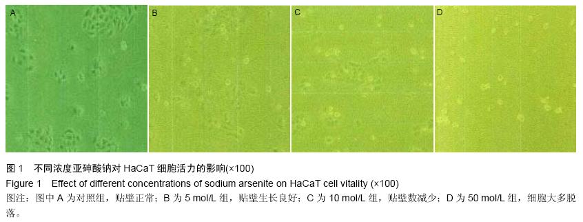
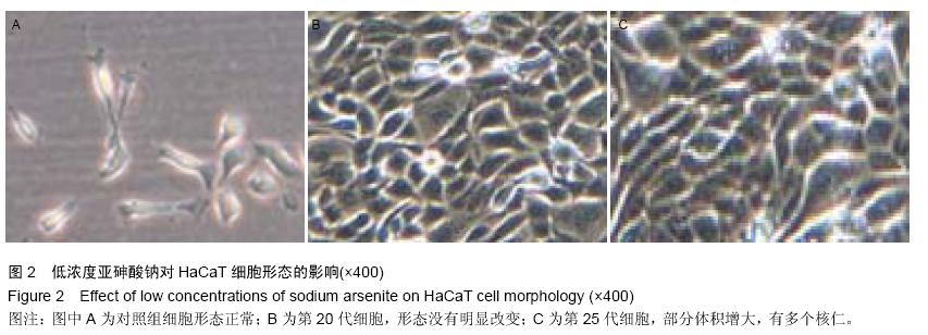
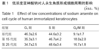
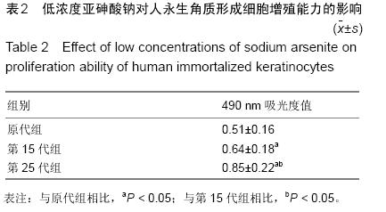
| [1] Liu X, Sun B, Wang X, et al. Synergistic effect of radon and sodium arsenite on DNA damage in HBE cells. Environ Toxicol Pharmacol. 2016;41:127-131.[2] Gutiérrez-Torres DS, González-Horta C, Del Razo LM, et al. Prenatal Exposure to Sodium Arsenite Alters Placental Glucose 1, 3, and 4 Transporters in Balb/c Mice. Biomed Res Int. 2015;2015:175025.[3] 张豫,罗鹏,叶建方,等.亚砷酸钠对人正常肝细胞凋亡及Bax和Bcl-2 mRNA和蛋白表达的影响[J].环境与健康杂志,2014,31(12):1048-1051.[4] Yao XF, Zheng BL, Bai J, et al. Low-level sodium arsenite induces apoptosis through inhibiting TrxR activity in pancreatic β-cells. Environ Toxicol Pharmacol. 2015;40(2):486-491. [5] Beevers C, Henderson D, Lillford L. et al. Investigation of sodium arsenite, thioacetamide, and diethanolamine in the alkaline comet assay: Part of the JaCVAM comet validation exercise. Mutat Res Genet Toxicol Environ Mutagen. 2015;786-788:165-171. [6] 刘世宜,胡楠楠,蔡思斯,等.亚砷酸钠对正常人类皮肤角质形成细胞蛋白激酶B及其下游信号因子1κB和NF-κB蛋白表达的影响[J].环境与健康杂志,2012,29(8):700-703.[7] Akinrinde AS, Olowu E, Oyagbemi AA, et al. Gastrointestinal protective efficacy of Kolaviron (a bi-flavonoid from Garcinia kola) following a single administration of sodium arsenite in rats: Biochemical and histopathological studies. Pharmacognosy Res. 2015;7(3):268-276.[8] George VC, Kumar DR, Suresh PK, et al. In Vitro Protective Potentials of Annona muricata Leaf Extracts Against Sodium Arsenite-induced Toxicity. Curr Drug Discov Technol. 2015;12(1):59-63.[9] Klibet F, Boumendjel A, Khiari M, et al. Oxidative stress-related liver dysfunction by sodium arsenite: Alleviation by Pistacia lentiscus oil. Pharm Biol. 2016; 54(2):354-363. [10] El-Jawahri AR, Vandusen HB, Traeger LN, et al. Quality of life and mood predict posttraumatic stress disorder after hematopoietic stem cell transplantation. Cancer. 2015. in press.[11] Dutta S, Krause A, Vosberg S, et al. The target cell of transformation is distinct from the leukemia stem cell in murine CALM/AF10 leukemia models. Leukemia. 2015. in press.[12] Zanoni TB, Hudari F, Munnia A, et al. The oxidation of p-phenylenediamine, an ingredient used for permanent hair dyeing purposes, leads to the formation of hydroxyl radicals: Oxidative stress and DNA damage in human immortalized keratinocytes. Toxicol Lett. 2015; 239(3):194-204. [13] Reijnders CM, van Lier A, Roffel S, et al. Development of a Full-Thickness Human Skin Equivalent In Vitro Model Derived from TERT-Immortalized Keratinocytes and Fibroblasts. Tissue Eng Part A. 2015;21(17-18): 2448-2459. [14] Arora S, Tyagi N, Bhardwaj A, et al. Silver nanoparticles protect human keratinocytes against UVB radiation-induced DNA damage and apoptosis: potential for prevention of skin carcinogenesis. Nanomedicine. 2015;11(5):1265-1275. [15] Di Grazia A, Cappiello F, Imanishi A, et al. The Frog Skin-Derived Antimicrobial Peptide Esculentin-1a (1-21)NH2 Promotes the Migration of Human HaCaT Keratinocytes in an EGF Receptor-Dependent Manner: A Novel Promoter of Human Skin Wound Healing? PLoS One. 2015;10(6):e0128663. [16] 金银龙,梁超轲,何公理,等.中国地方性砷中毒分布调查(总报告)[J].卫生研究,2003,32(6):519-540.[17] 高健伟,韦炳干,薛源,等.地方性砷中毒地区环境砷暴露健康风险研究进展[J].生态毒理学报,2013,8(2):138-147.[18] 赵曾艳.地方性砷中毒对机体氧化应激和免疫功能远期影响的研究[D].太原:山西医科大学,2010.[19] 郑艳艳,黄世文,姜岳明.不同暴露途径建立砷致癌动物模型研究的综述[J].工业卫生与职业病,2014,(3):221-224.[20] 闫婷.应用RDPCR检测职业砷暴露工人P53基因的损伤[D].昆明:昆明医科大学,2013.[21] 凌敏.ROS通过ERK/NF-kB调控miR-21在低水平亚砷酸钠所致细胞恶性转化中的作用[D].南京:南京医科大学, 2012.[22] 申璐.miR-21与MAPKs信号通路反馈调控在亚砷酸钠所致HELF细胞恶性转化中的作用[D].南京:南京医科大学, 2013.[23] 李静,吴顺华.砷致DNA甲基化变化与致癌作用的研究进展[J]. 国外医学(医学地理分册),2014,(3):247-251.[24] 李达圣, Kanehisa Morimoto,Tatsuya Takeshita,等.砷对人淋巴细胞DNA氧化损伤的作用[J].中华预防医学杂志, 2002,36(1):12-15.[25] Posey T, Weng T, Chen Z, et al. Arsenic-induced changes in the gene expression of lung epithelial L2 cells: implications in carcinogenesis. BMC Genomics. 2008;9:115.[26] Su PF, Hu YJ, Ho IC, et al. Distinct gene expression profiles in immortalized human urothelial cells exposed to inorganic arsenite and its methylated trivalent metabolites. Environ Health Perspect. 2006;114(3): 394-403.[27] Wang XJ, Yang J, Cang H, et al. Gene expression alteration during redox-dependent enhancement of arsenic cytotoxicity by emodin in HeLa cells. Cell Res. 2005;15(7):511-522.[28] Liu F, Jan KY. DNA damage in arsenite- and cadmium-treated bovine aortic endothelial cells. Free Radic Biol Med. 2000;28(1):55-63.[29] Takahashi M, Ota A, Karnan S, et al. Arsenic trioxide prevents nitric oxide production in lipopolysaccharide -stimulated RAW 264.7 by inhibiting a TRIF-dependent pathway. Cancer Sci. 2013;104(2):165-170.[30] Xia J, Li Y, Yang Q, et al. Arsenic trioxide inhibits cell growth and induces apoptosis through inactivation of notch signaling pathway in breast cancer. Int J Mol Sci. 2012;13(8):9627-9641. [31] Lau A, Whitman SA, Jaramillo MC, et al. Arsenic- mediated activation of the Nrf2-Keap1 antioxidant pathway. J Biochem Mol Toxicol. 2013;27(2):99-105.[32] Zhang Z, Wang X, Cheng S, et al. Reactive oxygen species mediate arsenic induced cell transformation and tumorigenesis through Wnt/β-catenin pathway in human colorectal adenocarcinoma DLD1 cells. Toxicol Appl Pharmacol. 2011;256(2):114-121.[33] Jomova K, Jenisova Z, Feszterova M, et al. Arsenic: toxicity, oxidative stress and human disease. J Appl Toxicol. 2011;31(2):95-107.[34] Platanias LC. Biological responses to arsenic compounds. J Biol Chem. 2009;284(28):18583-18587.[35] Liu Q, Zhang H, Smeester L, et al. The NRF2-mediated oxidative stress response pathway is associated with tumor cell resistance to arsenic trioxide across the NCI-60 panel. BMC Med Genomics. 2010; 3:37.[36] Mo J, Xia Y, Wade TJ, et al. Altered gene expression by low-dose arsenic exposure in humans and cultured cardiomyocytes: assessment by real-time PCR arrays. Int J Environ Res Public Health. 2011;8(6):2090-2108.[37] 肖骞.p53基因甲基化、DNMT3B基因多态性与燃煤型砷中毒关系研究[J].中国热带医学,2013,13(3):284-288.[38] 苏丽琴,金银龙.现场流行病学研究对慢性砷中毒判定的意义[J].卫生研究,2005,34(5):636-639.[39] 李远.NF-κB通过mot-2抑制p53在低水平亚砷酸钠所致细胞恶性转化中的作用[D].南京:南京医科大学,2013.[40] Han Y, Zhao H, Jiang Q, et al. Chemopreventive mechanism of polypeptides from Chlamy Farreri (PCF) against UVB-induced malignant transformation of HaCaT cells. Mutagenesis. 2015;30(2):287-296.[41] 梁文俊.三氧化二砷致HaCaT细胞恶性转化细胞的HSP27mRNA变化[D].大连:大连医科大学,2014. [42] Sun Y, Pi J, Wang X, et al. Aberrant cytokeratin expression during arsenic-induced acquired malignant phenotype in human HaCaT keratinocytes consistent with epidermal carcinogenesis. Toxicology. 2009; 262(2): 162-170.[43] Wang Q, Li WL, You P, et al. Oncoprotein BMI-1 induces the malignant transformation of HaCaT cells. J Cell Biochem. 2009;106(1):16-24.[44] 王大朋.Keap1-Nrf2/ARE 信号通路在砷致HaCaT 细胞恶性转化过程中的动态变化及其机制研究[D].苏州:苏州大学,2015.[45] Ren Q, Kari C, Quadros MR, et al. Malignant transformation of immortalized HaCaT keratinocytes through deregulated nuclear factor kappaB signaling. Cancer Res. 2006;66(10):5209-5215.[46] He YY, Pi J, Huang JL, et al. Chronic UVA irradiation of human HaCaT keratinocytes induces malignant transformation associated with acquired apoptotic resistance. Oncogene. 2006;25(26):3680-3688.[47] 蒙智娟,杨桥樱,黄世文,等.亚砷酸钠诱导人永生化表皮细胞恶性转化模型的建立[J].广西医学,2014,(5):561-564.[48] 周优优,高明阳.砷致HaCaT恶性转化细胞热休克蛋白27水平变化的观察[J].中国皮肤性病学杂志,2015,(2): 122-124,128.[49] Dedinszki D, Sipos A, Kiss A, et al. Protein phosphatase-1 is involved in the maintenance of normal homeostasis and in UVA irradiation-induced pathological alterations in HaCaT cells and in mouse skin. Biochim Biophys Acta. 2015;1852(1):22-33.[50] Vitale N, Kisslinger A, Paladino S, et al. Resveratrol couples apoptosis with autophagy in UVB-irradiated HaCaT cells. PLoS One. 2013;8(11):e80728. |
.jpg)