中国组织工程研究 ›› 2016, Vol. 20 ›› Issue (5): 628-634.doi: 10.3969/j.issn.2095-4344.2016.05.004

• 周围神经损伤动物模型 Animal models of peripheral nerve injury • 上一篇    下一篇

SD大鼠脊髓损伤后微环境的模拟实验

贾叙锋,龙 苗,戢 勇,黄光平,周 玉,张方德,冯大雄   

  1. 1简阳市人民医院骨科,四川省简阳市 641400;2泸州医学院附属医院脊柱外科,四川省泸州市 646000
  • 收稿日期:2015-11-03 出版日期:2016-01-29 发布日期:2016-01-29
  • 通讯作者: 冯大雄,博士, 教授,泸州医学院附属医院脊柱外科,四川省泸州市 646000
  • 作者简介:贾叙锋,男,1983年生,四川省安岳县人,2015年四川医科大学毕业,硕士,主治医师,主要从事脊髓损伤的研究。
  • 基金资助:
    国家自然科学基金资助(30872602)

Simulation of microenviroment after spinal cord injury in Sprague-Dawley rats

Jia Xu-feng1, 2, Long Miao1, Ji Yong1, Huang Guang-ping1, Zhou Yu1, Zhang Fang-de1, Feng Da-xiong2   

  1. 1Department of Orthopedics, People’s Hospital of Jianyang City, Jianyang 641400, Sichuan Province, China; 2Department of Spine Surgery, Affiliated Hospital of Luzhou Medical College, Luzhou 646000, Sichuan Province, China
  • Received:2015-11-03 Online:2016-01-29 Published:2016-01-29
  • Contact: Feng Da-xiong, M.D., Professor, Department of Spine Surgery, Affiliated Hospital of Luzhou Medical College, Luzhou 646000, Sichuan Province, China
  • About author:Jia Xu-feng, Master, Attending physician, Department of Orthopedics, People’s Hospital of Jianyang City, Jianyang 641400, Sichuan Province, China; Department of Spine Surgery, Affiliated Hospital of Luzhou Medical College, Luzhou 646000, Sichuan Province, China
  • Supported by:

    the National Natural Science Foundation of China, No. 30872602

摘要:

文章快速阅读:

文题释义:

脊髓损伤微环境改变:脊髓损伤后,早期病理变化体征为局部的水肿、结构破坏、神经元坏死以及对附件组织产生压迫加重了脊髓组织缺血损害等。在直接暴力损伤后出现的病理生理变化以及各种因素同样在脊髓损伤中起着极为重要的作用,如肿瘤坏死因子α、白细胞介素6等各种致炎细胞因子、炎性因子、炎性细胞活化等促使胶质细胞的大量增殖,并在病灶处形成致密的瘢痕组织,从而阻挡了神经纤维的延伸。

Allen’s脊髓损伤模型:采用改良Allen’s模型以改变重物下落的高度、致伤力,从而复制出不同类型、不同损伤程度的脊髓损伤后微环境。动物所受的致伤能量常以冲击棒质量(g)乘以坠落高度(cm)即克厘米势能(gcf)表示,致伤势能为20 g×5 cm,20 g×10 cm,20 g×15 cm,可模拟SD大鼠轻、中、重度脊髓损伤模型的微环境。


背景:通过制作SD大鼠的轻、中、重度脊髓损伤模型,使之符合基础实验研究的脊髓损伤类型,可进一步了解SD大鼠脊髓损伤后的微环境变化,为临床治疗提供帮助。
目的:模拟SD大鼠脊髓损伤后微环境,观察不同致伤势能的Allen’s模型和脊髓全切模型SD大鼠脊髓损伤后不同时间点的神经功能、病理以及运动诱发电位的变化。
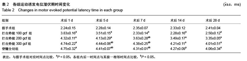
方法:选择125只成年雌性健康SD大鼠,随机分为假手术组、打击势能100 gcf组(20 g×5 cm)、打击势能200 gcf组(20 g×10 cm)、打击势能300 gcf组(20 g×15 cm)、脊髓全切组,每组25只,在制模成功后第1,5,7,14,28天采用BBB后肢运动功能评分、运动诱发电位监测、病理切片鉴定,观察各组大鼠脊髓损伤程度。
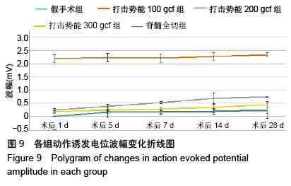
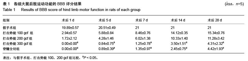
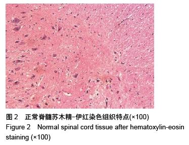
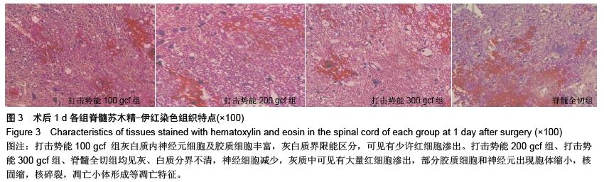
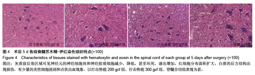
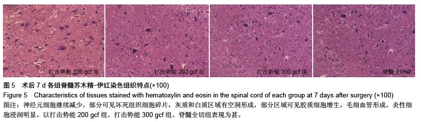
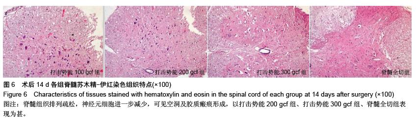
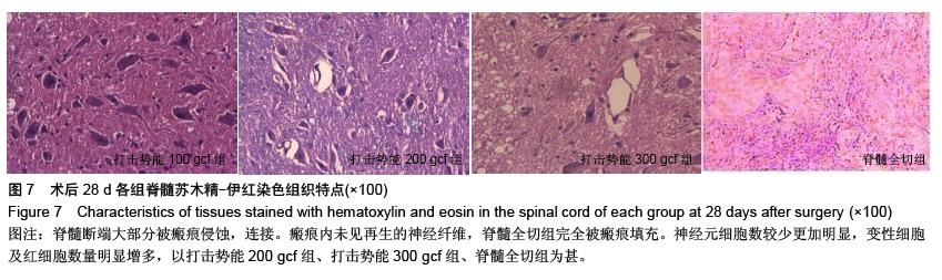
结果与结论:①整个实验中SD大鼠死亡24只,脊髓全切组的死亡率和并发症最高,每组的BBB评分均降低,随着时间延长BBB评分逐渐增高。各手术组与假手术组在对应时间点比较,差异有显著性意义(P < 0.05),打击势能300 gcf组和脊髓全切组在对应时间点比较,差异无显著性意义(P > 0.05)。②各手术组在伤后1 d病灶处出现炎性细胞浸润,神经元细胞肿胀明显,随着时间推移,神经细胞减少;伤后28 d发现大量星形胶质细胞增生,瘢痕及脊髓空洞形成,打击势能300 gcf组、脊髓全切组明显重于打击势能100 gcf 组、打击势能200 gcf组。③各手术组与假手术组的波幅及潜伏期在对应时间点比较,差异有显著性意义(P < 0.05);打击势能300 gcf组和脊髓全切组在对应时间点比较,差异无显著性意义(P > 0.05)。结果表明致伤势能为   20 g×5 cm,20 g×10 cm,20 g×15 cm,可模拟SD大鼠轻、中、重度脊髓损伤模型的微环境;不同势能的改良Allen’s模型较脊髓全切模型并发症低,更符合基础研究。 

ORCID:0000-0001-7648-8821(冯大雄)

关键词: 实验动物, 神经损伤与修复动物模型, 脊髓损伤, Allen’s模型, 脊髓全切模型, SD大鼠, 微环境, 国家自然科学基金

Abstract:

BACKGROUND: We built Sprague-Dawley rat models with mild, moderate, and severe spinal cord injuries to accord with the spinal cord injury types for basic empirical study, and consequently to further understand the microenvironmental change in Sprague-Dawley rats with spinal cord injury, and to provide help for clinical treatment.
OBJECTIVE: To observe the changes in nerve function, pathological manifestation and motor sensory evoked potential in Allen’s models and Sprague-Dawley rats with complete spinal cord transection at different time points after spinal cord injury by simulating the microenviroment in Sprague-Dawley rats.
METHODS: A total of 125 healthy adult female Sprague-Dawley rats were selected and randomly divided into group sham operation group, 100 gcf hit potential group (20 g×5 cm), 200 gcf hit potential (20 g×10 cm), 300 gcf hit potential group (20 g×15 cm), and spinal cord complete transection group with 25 rats in each group. At 1, 5, 7, 14 and 28 days after model establishment, the degree of spinal cord injury was identified by the BBB scores of motion function, motor evoked potential, and pathological section.
RESULTS AND CONCLUSION: (1) Totally 24 Sprague-Dawley rats died in the experiment. The death rate and the rate of complications were highest in the spinal cord complete transection group. The BBB score of each group was decreased. The BBB scores in every group increased as time went on. There were significant differences between each surgery group and the sham operation group at corresponding time points (P < 0.05).  No significant difference was found between the 300 gcf hit potential group and the spinal cord complete transection group at corresponding time points (P > 0.05). (2) In each surgery group, the infiltration of inflammatory cells and obvious swelling of neurons were visible at 1 day after injury. Neural cells reduced with time prolonged. At 28 days after injury, a large number of astrocytes proliferated, scar and spinal cord cavity formed. Above symptoms were worse in the 300 gcf hit potential group and spinal cord complete transection group than in the 100 gcf and 200 gcf hit potential groups. (3) Significant differences in amplitude and latency were detectable between each surgery group and the sham operation group (P < 0.05). No significant difference in amplitude and latency was detected between the 300 gcf hit potential group and the spinal cord complete transection group at corresponding time points (P > 0.05). Results confirmed that hit potential of 20 g×5 cm, 20 g×10 cm and 20 g×15 cm can simulate the microenvironment of Sprague-Dawley rats with mild, moderate and severe spinal cord injury. The rate of complication was lower in modified Allen’s model of different hit potentials than in models of spinal cord complete transection, and was more accorded with basic research.