中国组织工程研究 ›› 2015, Vol. 19 ›› Issue (20): 3124-3128.doi: 10.3969/j.issn.2095-4344.2015.20.002

• 骨组织构建 bone tissue construction • 上一篇    下一篇

退行性腰椎侧凸程度与骨质疏松症程度无关

彭毛加措1,高  强2   

  1. 1青海省人民医院骨科,青海省西宁市  810000;2解放军第九二医院,福建省南平市  353000
  • 出版日期:2015-05-14 发布日期:2015-05-14
  • 作者简介:彭毛加措,男,1974年生,青海省人,藏族,主治医师,主要从事关节与创伤的研究。

Degenerative lumbar scoliosis has no correlation with osteoporosis 

Pengmao Jiacuo1, Gao Qiang2   

  1. 1Department of Orthopedics, Qinghai Provincial People’s Hospital, Xining 810000, Qinghai Province, China; 2the 92nd Hospital of PLA, Nanping 353000, Fujian Province, China
  • Online:2015-05-14 Published:2015-05-14
  • About author:Pengmao Jiacuo, Attending physician, Department of Orthopedics, Qinghai Provincial People’s Hospital, Xining 810000, Qinghai Province, China

摘要:

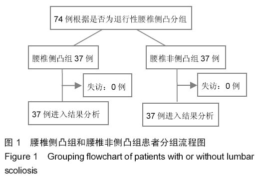
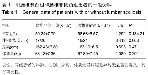
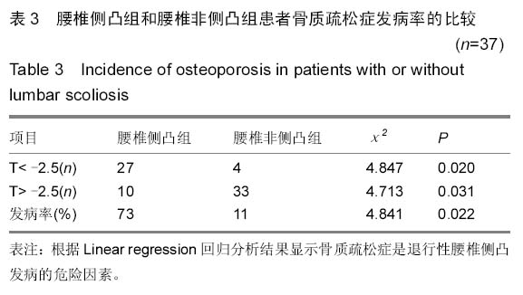
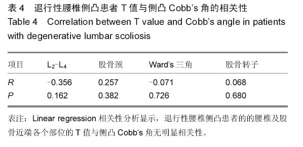
背景:骨质疏松是以全身骨组织显微结构损坏及全身骨量减少为特征,有学者认为骨质疏松患者腰椎椎体前高不会减少,反而会有增加的趋势。
目的:分析退行性腰椎侧凸与骨质疏松症的相关性。
方法:选取退行性腰椎侧凸患者37例,另选取同期在住院治疗的腰椎非侧凸组患者37例。测量时采用改良Cobb法,沿患者的T12-L5各椎体上终板平行划线,2条线的最大夹角为侧凸角,两线所在椎体即为上、下端椎。运用双能X射线吸收法对患者L2-L4、股骨颈、Ward’s三角区以及股骨转子的骨密度进行测定。采用Linear regression对骨质疏松和退行性腰椎侧凸症的相关性进行分析
结果与结论:腰椎侧凸组患者L2-L4、股骨颈、Ward’s三角区以及股骨转子部位的骨密度T值与腰椎非侧凸组比较差异有显著性意义(P < 0.05),其中股骨各部位骨密度较腰椎(L2-L4)较低。腰椎侧凸组患者骨质疏松症发病率明显高于腰椎非侧凸组患者(P < 0.05)。Linear regression回归分析结果显示骨质疏松症是退行性腰椎侧凸发病的危险因素;退行性腰椎侧凸患者的腰椎及股骨近端各个部位的T值与侧凸Cobb’s角无明显相关性。结果提示,骨质疏松症是退行性腰椎侧凸发病的危险因素,同时骨质疏松程度与侧凸程度无关。

关键词: 组织构建, 骨组织工程, 骨质疏松, 退行性腰椎侧凸, 骨密度, 相关性

Abstract:

BACKGROUND: Osteoporosis is characterized as bone microstructure damage of the whole body and systemic osteopenia. Some scholars believe that the anterior vertebral height of patients with osteoporosis cannot decrease, but shows a trend of increase. 
OBJECTIVE: To investigate the correlation between degenerative lumbar scoliosis and osteoporosis.
METHODS: Thirty-seven patients with degenerative lumbar scoliosis were selected as observation group and another 37 inpatients with no lumbar scoliosis as control group. Modified Cobb method was used to detect the maximum angle between parallel lines to the endplate on the T12-L5 vertebrae, and the vertebrae with these two parallel lines were regarded as upper and lower vertebrae. Dual energy X-ray absorptiometry was used to measure the bone mineral density of L2-L4 segments, femoral neck, Ward’s triangle and femoral trochanter. Linear regression analysis was used to analyze the correlation between osteoporosis and degenerative lumbar scoliosis.
RESULTS AND CONCLUSION: There were significant differences in the T values in the L2-L4 segments, femoral neck, Ward’s triangle and femoral trochanter between the observation and control groups (P < 0.05), and the bone density of the femoral parts was lower than that of the lumbar vertebrae (L2-L4). The incidence of osteoporosis in the patients with lumbar scoliosis was significantly higher than that in the patients with no lumbar scoliosis (P < 0.05). Osteoporosis was a risk factor for degenerative lumbar scoliosis, and the T values of the lumbar vertebra and proximal femur had no remarkable correlation with Cobb’s angles. These findings indicate that osteoporosis is the risk factor for degenerative lumbar scoliosis, but it has no correlation with the severity of scoliosis.

Key words: Osteoporosis, Scoliosis, Bone Density

中图分类号: