中国组织工程研究 ›› 2022, Vol. 26 ›› Issue (17): 2685-2689.doi: 10.12307/2022.535

• 组织构建实验造模 experimental modeling in tissue construction • 上一篇    下一篇

诱导巨噬细胞向M2极化降低糖尿病视网膜病变模型小鼠的氧化损伤

殷  亮,张明雪,李家男,江  枫   

  1. 天津医科大学总医院,天津市   300014
  • 收稿日期:2021-06-15 修回日期:2021-06-16 接受日期:2021-07-24 出版日期:2022-06-18 发布日期:2021-12-24
  • 通讯作者: 殷亮,硕士,主治医师,天津医科大学总医院,天津市 300014
  • 作者简介:殷亮,男,1980年生,辽宁省人,满族,2009年辽宁医学院毕业,硕士,主治医师,主要从事角膜病、白内障诊治的研究。
  • 基金资助:
    国家自然科学基金资助项目(81800853),项目负责人:江枫

Inducing macrophage polarization to M2 anti-inflammatory type reduces oxidative damage in diabetic retinopathy mice

Yin Liang, Zhang Mingxue, Li Jianan, Jiang Feng   

  1. General Hospital of Tianjin Medical University, Tianjin 300014, China
  • Received:2021-06-15 Revised:2021-06-16 Accepted:2021-07-24 Online:2022-06-18 Published:2021-12-24
  • Contact: Yin Liang, General Hospital of Tianjin Medical University, Tianjin 300014, China
  • About author:Yin Liang, Master, Attending physician, General Hospital of Tianjin Medical University, Tianjin 300014, China
  • Supported by:
    the National Natural Science Foundation of China, No. 81800853 (to JF)

摘要:

文题释义:
TNFSF15:肿瘤坏死因子超家族,血管内皮生长抑制因子,由正常的血管内皮细胞分泌,其可显著抑制新生血管的生成,同时其可以抑制内皮细胞进入有丝分裂,并诱导增殖状态的内皮细胞发生程序性凋亡。

背景:糖尿病视网膜病变的主要标志之一就是新生血管的持续生成,而TNFSF15作为血管生成抑制因子可显著抑制血管生成。
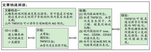
目的:探究TNFSF15通过诱导巨噬细胞向M2极化降低糖尿病视网膜病变小鼠氧化损伤的作用机制。
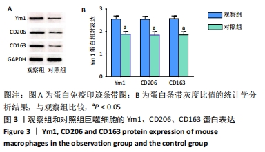
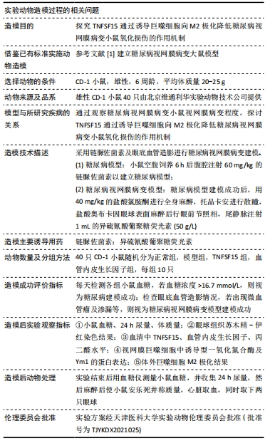
方法:选取40只雄性CD-1小鼠,随机分为正常组、模型组、TNFSF15组和血管内皮生长因子组,每组10只。除正常组外,其他各组小鼠采用链脲佐菌素及眼底血管造影进行糖尿病视网膜病变建模。建模成功后,TNFSF15组小鼠腹腔内注射250 mg/L的TNFSF15;血管内皮生长因子组小鼠腹腔内注射250 mg/L的血管内皮生长因子;正常组和模型组同期灌胃同体积生理盐水。实验完成后,取小鼠视网膜组织,提取巨噬细胞进行体外培养,分为2组,加入TNFSF15的为观察组及加入 PBS的为对照组。苏木精-伊红染色检测小鼠网膜病理形态;ELISA法检测血清中TNFSF15、血管内皮生长因子、丙二醛、超氧化物歧化酶水平;Western blot检测小鼠视网膜组织中诱导型一氧化氮合酶、Ym1、CD206、CD163的蛋白表达,并观察体外巨噬细胞M2极化结果。实验方案经天津医科大学实验动物伦理委员会批准(批准号为TJYKDX2021025)。
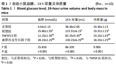
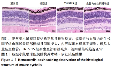
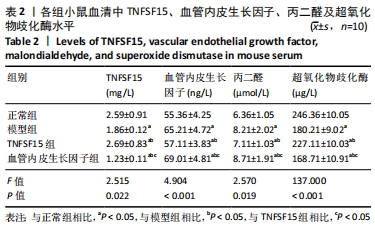
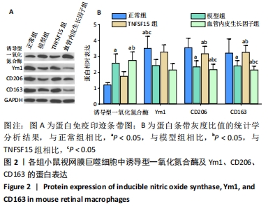
结果与结论:①与正常组比较,模型组血糖及24 h尿量、血清血管内皮生长因子及丙二醛水平、视网膜巨噬细胞中诱导型一氧化氮合酶蛋白表达明显升高(P < 0.05),上述指标TNFSF15组较模型组明显降低(P < 0.05),血管内皮生长因子组较TNFSF15组明显升高(P < 0.05);与正常组比较,模型组体质量、血清中TNFSF15及超氧化物歧化酶水平、Ym1、CD206及CD163蛋白表达明显降低(P < 0.05),上述指标TNFSF15组较模型组明显升高(P < 0.05),血管内皮生长因子组较TNFSF15组明显降低(P < 0.05);②正常组小鼠视网膜结构正常且排列整齐;模型组与血管内皮生长因子组出现明显病变;TNFSF15组与前两组相比,其新生血管明显减少,视网膜结构趋近正常;③细胞实验中,与对照组相比,观察组的Ym1、CD206及CD163的蛋白表达显著增加(P < 0.05);④结论:TNFSF15可能是通过诱导巨噬细胞向M2极化,来降低糖尿病视网膜病变小鼠的氧化损伤,改善视网膜病变程度。

https://orcid.org/0000-0001-8848-7375 (殷亮) 

中国组织工程研究杂志出版内容重点:组织构建;骨细胞;软骨细胞;细胞培养;成纤维细胞;血管内皮细胞;骨质疏松;组织工程

关键词: TNFSF15, 巨噬细胞, M2极化, 糖尿病视网膜病变, 氧化损伤, 小鼠

Abstract: BACKGROUND: One of the main signs in diabetic retinopathy is the continuous formation of new blood vessels, and tumor necrosis factor ligand superfamily member 15 (TNFSF15), as an angiogenesis inhibitor, can significantly inhibit angiogenesis.
OBJECTIVE: To investigate the mechanism by which TNFSF15 reduces oxidative damage in diabetic retinopathy mice by inducing macrophage polarization to M2 anti-inflammatory type. 
METHODS: Forty male CD-1 mice were randomly divided into four groups (n=10 per group): a normal group, a model group, a TNFSF15 group, and a vascular endothelial growth factor group. Except for the normal group, a diabetic retinopathy model was established in all the groups by using streptozotocin and fundus angiography respectively. After the success of the modeling, mice in the TNFSF15 group were intraperitoneally injected with 250 mg/L TNFSF15, and mice in the vascular endothelial growth factor group were intraperitoneally injected with 250 mg/L vascular endothelial growth factor. Mice in the normal group and the model group were given the same volume of normal saline simultaneously. After the experiment was completed, the mouse retina was taken, and macrophages were extracted for in vitro culture. The cells were cultured with TNFSF15 in observation group or with phosphate buffered saline in control group. Hematoxylin-eosin staining was used to detect the pathological morphology of mouse omentum. Enzyme linked immunosorbent assay was used to detect serum levels of TNFSF15, vascular endothelial growth factor, malondialdehyde, and superoxide dismutase. Western blot was used to detect the protein expression of inducible nitric oxide synthase, Ym1, CD206, and CD163 in mouse retinal tissue. In vitro macrophage polarization to M2 anti-inflammatory type was also observed. This study protocol was approved by the Experimental Animal Ethics Committee of Tianjin Medical University (approval No. TJYKDX2021025).
RESULTS AND CONCLUSION: Compared with the normal group, the blood glucose level and 24-hour urine volume, the serum level of vascular endothelial growth factor and malondialdehyde, and the protein expression of inducible nitric oxide synthase in retinal macrophages were significantly increased in model group (P < 0.05). While the above indicators in the TNFSF15 group were significantly lower than those in the model group (P < 0.05), and those in the vascular endothelial growth factor group were significantly higher than those in the TNFSF15 group (P < 0.05). Compared with the normal group, the body mass, serum levels of TNFSF15 and superoxide dismutase, and protein expression of YM1, CD206 and CD163 were significantly decreased in the model group (P < 0.05), while the above indicators in the TNFSG15 group were significantly higher than those in the model group (P < 0.05), and these indicators in the vascular endothelial growth factor group were significantly lower than those in the TNFSF15 group (P < 0.05). The mouse retina in the normal group had normal structure that was in alignment, and obvious retinal lesions were observed in the model group and vascular endothelial growth factor group. Compared with the model group and vascular endothelial growth factor group, there were fewer new blood vessels in the TNFSR15 group and its retinal structure intended to be normal. In the cell experiment, the protein expression of Ym1, CD206, and CD163 was increased significantly in the observation group compared with the control group (P < 0.05). Therefore, TNFSF15 may reduce oxidative damage in diabetic retinopathy mice and improve retinopathy by inducing macrophage polarization to M2 anti-inflammatory type.

Key words: tumor necrosis factor ligand superfamily member 15, macrophage, M2 polarization, diabetic retinopathy, oxidative damage, mouse

中图分类号: