中国组织工程研究 ›› 2017, Vol. 21 ›› Issue (4): 564-573.doi: 10.3969/j.issn.2095-4344.2017.04.012
• 组织构建实验造模 experimental modeling in tissue construction • 上一篇 下一篇
两种方法建立兔腰椎间盘退变模型
孟祥玉,巴穆登,艾尔肯•阿木冬
- 新疆医科大学第六附属医院脊柱外科,新疆维吾尔自治区乌鲁木齐市 830000
Establishment of a rabbit model of intervertebral disc degeneration using two methods
Meng Xiang-yu, Ba Mu-deng, Aierken•Amudong
- Department of Spine Surgery, the Sixth Affiliated Hospital of Xinjiang Medical University, Urumqi 830000, Xinjiang Uygur Autonomous Region, China
摘要:
文章快速阅读:
.jpg) 文题释义:
髓核抽吸法建立椎间盘退变模型:用16 G注射针头在椎间隙平行终板方向刺入5.0-6.0 mm,持续负压吸引10 s。髓核抽吸法作为目前较成熟的造模方法,诱导椎间盘退变效果明确,退变程度缓和。
纤维环薄层切除法建立椎间盘退变模型:显微镜辅助下用单刃刀在纤维环表面切除1 mm×2 mm范围,约占纤维环层厚的1/2(约1 mm),避免切透纤维环造成髓核溢出。纤维环薄层切除法直接使纤维环结构受到不完全破裂,造成髓核内环境变化,髓核内蛋白多糖水分丢失,明确导致椎间盘发生退变,进一步佐证了人类纤维环结构的老化、结构不完整造成的椎间盘退变假说。
文题释义:
髓核抽吸法建立椎间盘退变模型:用16 G注射针头在椎间隙平行终板方向刺入5.0-6.0 mm,持续负压吸引10 s。髓核抽吸法作为目前较成熟的造模方法,诱导椎间盘退变效果明确,退变程度缓和。
纤维环薄层切除法建立椎间盘退变模型:显微镜辅助下用单刃刀在纤维环表面切除1 mm×2 mm范围,约占纤维环层厚的1/2(约1 mm),避免切透纤维环造成髓核溢出。纤维环薄层切除法直接使纤维环结构受到不完全破裂,造成髓核内环境变化,髓核内蛋白多糖水分丢失,明确导致椎间盘发生退变,进一步佐证了人类纤维环结构的老化、结构不完整造成的椎间盘退变假说。
文题释义:
髓核抽吸法建立椎间盘退变模型:用16 G注射针头在椎间隙平行终板方向刺入5.0-6.0 mm,持续负压吸引10 s。髓核抽吸法作为目前较成熟的造模方法,诱导椎间盘退变效果明确,退变程度缓和。
纤维环薄层切除法建立椎间盘退变模型:显微镜辅助下用单刃刀在纤维环表面切除1 mm×2 mm范围,约占纤维环层厚的1/2(约1 mm),避免切透纤维环造成髓核溢出。纤维环薄层切除法直接使纤维环结构受到不完全破裂,造成髓核内环境变化,髓核内蛋白多糖水分丢失,明确导致椎间盘发生退变,进一步佐证了人类纤维环结构的老化、结构不完整造成的椎间盘退变假说。摘要
背景:国内外学者对椎间盘退变动物模型的建立做了大量工作,通过改变椎间盘力学特性、纤维环损伤、酶化学法等多种建模方式模拟椎间盘退变的病理表现。
目的:通过髓核抽吸法和纤维环薄层切除法建立椎间盘退变模型,比较两种方法的造模效果。
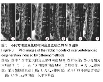
方法:取6月龄新西兰大白兔20只,通过手术暴露L3-4、L4-5及L5-6椎间盘,随机使用髓核抽吸法和纤维环薄层切除法对L3-4、L4-5椎间盘进行干预:①髓核抽吸组——利用16 G针头在椎间盘纤维环穿刺并负压抽吸10 s;②纤维环薄层切除组——在显微镜下辅助下,在兔椎间盘纤维环上切除1 mm×2 mm范围的约1/2层纤维环组织(避免切透全层)。于造模第1,3,5,9,13周进行核磁共振、X射线检查及椎间盘Masson染色病理学观察。
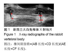
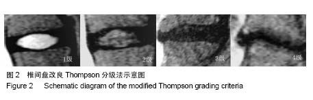
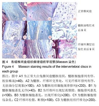
结果与结论:①X射线检查:纤维环薄层切除组椎间隙高度自术后第1周起便发生不同程度降低;髓核抽吸组椎间隙在术后1周及3周轻微降低,自第5周起发生较明显降低;纤维环薄层切除组术后第3,5,9周椎间隙高度指数百分比高于髓核抽吸组(P < 0.05);②核磁共振检查:两种方法均诱导相应椎间盘发生退变,各组随时间推移椎间盘退变程度逐渐加重,并于9周后均达到4级退变;髓核抽吸组术后1,3,5周的椎间盘退变程度重于纤维环薄层切除组(P < 0.05);③病理学观察:随时间推移,两组椎间盘髓核及纤维环退变程度逐渐加重;④结果表明:髓核抽吸法和纤维环薄层切除法均能诱导椎间盘退变,纤维环薄层切除法在5周内造成的椎间盘退变程度较髓核抽吸法轻,纤维环薄层切除法在纤维环上所造成的标示部位可作为纤维环修补研究的靶点。
中国组织工程研究杂志出版内容重点:组织构建;骨细胞;软骨细胞;细胞培养;成纤维细胞;血管内皮细胞;骨质疏松;组织工程
ORCID: 0000-0002-8143-522X(艾尔肯•阿木冬)
中图分类号:

.jpg) 文题释义:
髓核抽吸法建立椎间盘退变模型:用16 G注射针头在椎间隙平行终板方向刺入5.0-6.0 mm,持续负压吸引10 s。髓核抽吸法作为目前较成熟的造模方法,诱导椎间盘退变效果明确,退变程度缓和。
纤维环薄层切除法建立椎间盘退变模型:显微镜辅助下用单刃刀在纤维环表面切除1 mm×2 mm范围,约占纤维环层厚的1/2(约1 mm),避免切透纤维环造成髓核溢出。纤维环薄层切除法直接使纤维环结构受到不完全破裂,造成髓核内环境变化,髓核内蛋白多糖水分丢失,明确导致椎间盘发生退变,进一步佐证了人类纤维环结构的老化、结构不完整造成的椎间盘退变假说。
文题释义:
髓核抽吸法建立椎间盘退变模型:用16 G注射针头在椎间隙平行终板方向刺入5.0-6.0 mm,持续负压吸引10 s。髓核抽吸法作为目前较成熟的造模方法,诱导椎间盘退变效果明确,退变程度缓和。
纤维环薄层切除法建立椎间盘退变模型:显微镜辅助下用单刃刀在纤维环表面切除1 mm×2 mm范围,约占纤维环层厚的1/2(约1 mm),避免切透纤维环造成髓核溢出。纤维环薄层切除法直接使纤维环结构受到不完全破裂,造成髓核内环境变化,髓核内蛋白多糖水分丢失,明确导致椎间盘发生退变,进一步佐证了人类纤维环结构的老化、结构不完整造成的椎间盘退变假说。