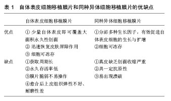
| [1] Pickerill HP. On the possibility of establishing skin banks. Br J Phst Surg. 1951;(4):157-165.[2] Reinwald JG, Green H. Serial cultivation of strains of human epidermal keratinocytes: the formation of keratinizing colonies from single cells. Cell. 1975;6(3):331-343.[3] Vig K, Chaudhari A, Tripathi S, et al. Advances in Skin Regeneration Using Tissue Engineering. Int J Mol Sci. 2017. doi: 10.3390/ijms18040789.[4] Marino D, Reichmann E, Meuli M. Skingineering. Eur J Pediatr Surg. 2014;24(3):205-213. [5] Chua AW, Khoo YC, Tan BK, et al. Skin tissue engineering advances in severe burns: review and therapeutic applications. Burns Trauma. 2016; 4:3. [6] Bello YM, Falabella AF. Use of skin substitutes in dermatology. Dermatol Clin. 2001;19:555-561.[7] Nyame TT, Chiang HA, Leavitt T, et al. Tissue-Engineered Skin Substitutes. Plast Reconstr Surg. 2015;136(6): 1379-1388.[8] Varkey M, Ding J, Tredget EE. Advances in Skin Substitutes-Potential of Tissue Engineered Skin for Facilitating Anti-Fibrotic Healing. J Funct Biomater. 2015; 6(3):547-563. [9] Markeson D, Pleat JM, Sharpe JR, et al. Scarring, stem cells, scaffolds and skin repair. J Tissue Eng Regen Med. 2015; 9(6):649-668. [10] Catalano E, Cochis A, Varoni E, et al. Tissue-engineered skin substitutes: an overview. J Artif Organs. 2013;16(4):397-403. [11] Kitala D, Kawecki M, Klama-Bary?a A, et al. Autologous skin grafts in the therapy of patients with burn injuries: a restrospective, open-label clinical study with pair matching. Adv Clin Exp Med. 2016;25(5):923-929. [12] Nyame TT, Chiang HA, Orgill DP. Clinical applications of skin substitutes. Surg Clin North Am. 2014;94(4):839-850. [13] Límová M. Active wound coverings: bioengineered skin and dermal substitutes. Surg Clin North Am. 2010;90(6): 1237-1255.[14] Van Der Veen VC, van der Wal MBA, van Leuwen MCE, et al. Biological background of dermal substitutes. Burns. 2010;36: 305-321.[15] Murray RC, Gordin EA, Saigal K, et al. Reconstruction of the radial forearm free flap donor site using Integra artificial dermis. Microsurgery. 2011;31:104-108.[16] Nguyen DQ, Potokar TS, Price P. An objective long-term evaluation of Integra (a dermal skin substitute) and split thickness skin grafts, in acute burns and reconstructive surgery. Burns. 2010;36:23-28.[17] Rnjak J, Wise SG, Mithieux SM, et al. Severe burn injuries and the role of elastin in the design of dermal substitutes. Tissue Eng. 2010;17(2):81-93.[18] Haslik W, Kamolz LP, Manna F et al. Management of full-thickness skin defects in the hand and wrist region: first long-term experiences with the dermal matrix matriderm. J Plast Reconstr Aesthet Surg. 2010;63:360-364.[19] Halim AS, Khoo TL, Mohd Yussof SJ. Biologic and synthetic skin substitutes: an overview. Indian J Plast Surg. 2010;43: S23-S28. [20] Varkey M, Ding J, Tredget EE. Advances in Skin Substitutes-Potential of Tissue Engineered Skin for Facilitating Anti-Fibrotic Healing. J Funct Biomater. 2015:6(3): 547-563. [21] Watt FM,Hogan BL. Out of Eden:Stem cells and their niches. Science. 2000:25;287(5457):1427-1430.[22] Nishimura E, Siobhan A, Oshima JH, et al. Dominant role of the niche in melanoeyte stem-cell fate determination. Nature. 2002;25;416(6883):854-860.[23] Xie T, Spradling AC. A niche maintaining germ line stem cells in the drosophila ovary. Science. 2000;13;290(5490): 328-330.[24] Ideta R, Soma T, Tsunenaga M, et al. Cultured human dermal papilla cells secrete a chemotactic factor for melanoeytes. J Dermatol Sci. 2002;28(1):48-59.[25] Gragnani A, Morgan JR, Ferreira LM. Differentiation and barrier formation of a cultured composite skin graft. J Burn Care Rehabil. 2002;23(2):126-131.[26] Wencel A, Zakrzewska KE, Samluk A, et al. Dried human skin fibroblasts as a new substratum for functional culture of hepatic cells. Acta Biochim Pol. 2017;64(2):357-363.[27] Koch M, Ehrenreich T, Koehl G, et al. Do cell based Tissue Engineering products for meniscus regeneration influence vascularization? Clin Hemorheol Microcirc. 2017. doi: 10.3233/CH-17085. [28] Wang JP, Zhou XL, Yan JP, et al. Nanobubbles as ultrasound contrast agent for facilitating small cell lung cancer imaging. Oncotarget. 2017. doi: 10.18632/oncotarget.18155.[29] Abaci HE, Guo Z, Doucet Y, et al. Next generation human skin constructs as advanced tools for drug development. Exp Biol Med (Maywood). 2017. doi: 10.1177/1535370217712690.[30] Pielesz A, Binia? D, Bobiński R, et al. The role of topically applied l-ascorbic acid in ex-vivo examination of burn-injured human skin. Spectrochim Acta A Mol Biomol Spectrosc. 2017. doi: 10.1016/j.saa.2017.05.055.[31] Modena DAO, da Silva CN Pt, Grecco C, et al. Extracorporeal shockwave: mechanisms of action and physiological aspects for cellulite, body shaping and localized fat - systematic review. J Cosmet Laser Ther. 2017. doi: 10.1080/14764172.2017. 1334928. [32] Kühbacher A, Burger-Kentischer A, Rupp S. Interaction of Candida Species with the Skin.Microorganisms. 2017. doi: 10.3390/microorganisms5020032. [33] Khan TK, Wender PA, Alkon DL. Bryostatin and its synthetic analog, picolog rescue dermal fibroblasts from prolonged stress and contribute to survival and rejuvenation of human skin equivalents. J Cell Physiol. 2017. doi: 10.1002/jcp.26043. [34] Szlavicz E, Szabo K, Groma G, et al. Splicing factors differentially expressed in psoriasis alter mRNA maturation of disease-associated EDA+ fibronectin. Mol Cell Biochem. 2017. doi: 10.1007/s11010-017-3090-1.[35] Kim BS, Lee JS, Gao G, et al. Direct 3D cell-printing of human skin with functional transwell system. Biofabrication. 2017; 9(2):025034. [36] Desmet E, Ramadhas A, Lambert J, et al. In vitro psoriasis models with focus on reconstructed skin models as promising tools in psoriasis research. Exp Biol Med (Maywood). 2017. doi: 10.1177/1535370217710637.[37] Henderson F, Alho R, Riches P, et al. Assessment of knee alignment with varus and valgus force through the range of flexion with non-invasive navigation. J Med Eng Technol. 2017. doi: 10.1080/03091902.2017.1333164.[38] Hu B, Leow WR, Amini S, et al. Orientational Coupling Locally Orchestrates a Cell Migration Pattern for Re-Epithelialization. Adv Mater. 2017. doi: 10.1002/adma.201700145. [39] Cai M, Shen S, Li H. The effect of facial expressions on respirators contact pressures. Comput Methods Biomech Biomed Engin. 2017. doi: 10.1080/10255842.2017.1336549. [40] Wang XR, Gao SQ, Niu XQ, et al. Capsaicin-loaded nanolipoidal carriers for topical application: design, characterization, and in vitro/in vivo evaluation. Int J Nanomedicine. 2017. doi: 10.2147/IJN.S131901.[41] Ma J, Chen B, Zhang Y, et al. Multiple laser pulses in conjunction with an optical clearing agent to improve the curative effect of cutaneous vascular lesions. Lasers Med Sci. 2017. doi: 10.1007/s10103-017-2244-4. [42] Strong AL, Neumeister MW, Levi B. Stem Cells and Tissue Engineering: regeneration of the Skin and Its Contents. Clin Plast Surg. 2017. doi: 10.1016/j.cps.2017.02.020. [43] Carvalho IC, Mansur HS. Engineered 3D-scaffolds of photocrosslinked chitosan-gelatin hydrogel hybrids for chronic wound dressings and regeneration. Mater Sci Eng C Mater Biol Appl. 2017. doi: 10.1016/j.msec.2017.04.126.[44] Beiki B, Zeynali B, Seyedjafari E. Fabrication of a three dimensional spongy scaffold using human Wharton's jelly derived extra cellular matrix for wound healing. Mater Sci Eng C Mater Biol Appl. 2017. doi: 10.1016/j.msec.2017.04.074. [45] Tan J, Zhao C, Zhou J, et al. Co-culturing epidermal keratinocytes and dermal fibroblasts on nano-structured titanium surfaces. Mater Sci Eng C Mater Biol Appl. 2017. doi: 10.1016/j.msec.2017.04.036. [46] Mostmans Y, Cutolo M, Giddelo C, et al. The role of endothelial cells in the vasculopathy of systemic sclerosis: A systematic review: SSc vasculopathy: The role of endothelial cells. Autoimmun Rev. 2017. doi: 10.1016/j.autrev.2017.05.024.[47] Chen X, Zhang M, Wang X, et al. Peptide-modified chitosan hydrogels promote skin wound healing by enhancing wound angiogenesis and inhibiting inflammation. Am J Transl Res. 2017;9(5):2352-2362. [48] Lee MJ, Hung SH, Huang MC, et al. Doxycycline potentiates antitumor effect of 5-aminolevulinic acid-mediated photodynamic therapy in malignant peripheral nerve sheath tumor cells. PLoS One. 2017. doi: 10.1371/journal.pone.0178493.[49] Cortese FAB, Aguiar S, Santostasi G. Induced Cell Turnover: a novel therapeutic modality for in situ tissue regeneration. Hum Gene Ther. 2017. doi: 10.1089/hum.2016.167. [50] Lotz C, Schmid FF, Oechsle E, et al. Cross-linked Collagen Hydrogel Matrix Resisting Contraction To Facilitate Full-Thickness Skin Equivalents. ACS Appl Mater Interfaces. 2017. doi: 10.1021/acsami.7b04017. [51] Mohafez H, Ahmad SA, Hadizadeh M, et al. Quantitative assessment of wound healing using high-frequency ultrasound image analysis. Skin Res Technol. 2017. doi: 10.1111/srt.12388.[52] Bhardwaj N, Chouhan D, Mandal BB. Tissue engineered skin and wound healing: current strategies and future directions. Curr Pharm Des. 2017. doi: 10.2174/1381612823666170526094606. [53] von Byern J, Mebs D, Heiss E, et al. Salamanders on the bench - A biocompatibility study of salamander skin secretions in cell cultures. Toxicon. 2017. doi: 10.1016/j.toxicon.2017.05.021. [54] Bonnet V, Richard V, Camomilla V, et al. Joint kinematics estimation using a multi-body kinematics optimisation and an extended Kalman filter, and embedding a soft tissue artefact model. J Biomech. 2017. doi: 10.1016/j.jbiomech.2017.04.033. [55] Aurand AM, Dufour JS, Marras WS. Accuracy map of an optical motion capture system with 42 or 21 cameras in a large measurement volume. J Biomech. 2017. doi: 10.1016/j.jbiomech.2017.05.006. [56] Fong CJ, Garzon MC, Hoi JW, et al. Assessment of Infantile Hemangiomas Using a Handheld Wireless Diffuse Optical Spectroscopic Device. Pediatr Dermatol. 2017. doi: 10.1111/pde.13150. [57] van den Broek LJ, Bergers LIJC, Reijnders CMA, et al. Progress and Future Prospectives in Skin-on-Chip Development with Emphasis on the use of Different Cell Types and Technical Challenges. Stem Cell Rev. 2017. doi: 10.1007/s12015-017-9737-1.[58] Sasaki Y, Sathi GA, Yamamoto O. Wound healing effect of bioactive ion released from Mg-smectite. Mater Sci Eng C Mater Biol Appl. 2017. doi: 10.1016/j.msec.2017.03.236.[59] Levengood SL, Erickson AE, Chang FC, et al. Chitosan-Poly(caprolactone) Nanofibers for Skin Repair. J Mater Chem B Mater Biol Med. 2017. doi: 10.1039/C6TB03223K. [60] Wang H, Dong XX, Yang JC, et al. Finite element method simulating temperature distribution in skin induced by 980-nm pulsed laser based on pain stimulation. Lasers Med Sci. 2017. doi: 10.1007/s10103-017-2223-9. [61] Scalise A, Torresetti M, Verdini F, et al. Acellular dermal matrix and heel reconstruction: a new prospective. J Appl Biomater Funct Mater. 2017. doi: 10.5301/jabfm.5000357. [62] Yu K, Lu F, Li Q, et al. In situ assembly of Ag nanoparticles (AgNPs) on porous silkworm cocoon-based would film: enhanced antimicrobial and wound healing activity. Sci Rep. 2017 May 18;7(1):2107. doi: 10.1038/s41598-017-02270-6.[63] Motamed S, Taghiabadi E, Molaei H, et al. Cell-based skin substitutes accelerate regeneration of extensive burn wounds in rats. Am J Surg. 2017. doi: 10.1016/j.amjsurg.2017.04.010. [64] Chang CJ, Yu DY, Hsiao YC, et al. Noninvasive imaging analysis of biological tissue associated with laser thermal injury. Biomed J. 2017. doi: 10.1016/j.bj.2016.10.004.[65] Chen S, Liu B, Carlson MA, et al. Recent advances in electrospun nanofibers for wound healing. Nanomedicine (Lond). 2017. doi: 10.2217/nnm-2017-0017. |
.jpg)

.jpg)