| [1]Khan Y, Yaszemski MJ, Mikos AG, Laurencin CT. Tissue engineering of bone: material and matrix considerations. J Bone Joint Surg Am. 2008;90 Suppl 1:36-42.
[2]Laurie SW, Kaban LB, Mulliken JB, et al. Donor-site morbidity after harvesting rib and iliac bone. Plastic and reconstructive surgery. 1984;73:933-938.
[3]韩冰,付小兵.间充质干细胞的研究进展与临床应用前景[J].中国修复重建外科杂志, 2006,20(12):1257-1261.
[4]马锡慧,冯凯,石炳毅.人脐带间充质干细胞生物学特性及其研究进展[J].中国组织工程研究, 2011,15(32): 6064-6067.
[5]Zvaifler NJ, Marinova-Mutafchieva L, Adams G, et al. Mesenchymal precursor cells in the blood of normal individuals. Arthritis Res. 2000;2(6):477-488.
[6]Deb A, Wang S, Skelding KA, et al. Bone marrow-derived cardiomyocytes are present in adult human heart: a study of gender-mismatched bone marrow transplantation patients. Circulation. 2003; 107(9):1247-1249.
[7]Guilak F, Lott KE, Awad HA, et al. Clonal analysis of the differentiation potential of human adipose-derived adult stem cells. J Cell Physiol. 2006;206(1):229-237.
[8]Li S, Huang KJ, Wu JC, et al. Peripheral blood-derived mesenchymal stem cells: candidate cells responsible for healing critical-sized calvarial bone defects. Stem Cells Transl Med. 2015;4(4):359-68.
[9]杨志明.组织工程骨的研究成果及存在的问题[J].中国修复重建外科杂志,2005,19(2):87-89.
[10]贝抗胜,吴礼杨,孙庆文,等.人骨膜细胞生物学特性的实验研究[J].中华创伤骨科杂志,2011,13(12):1170-1174.
[11]张大伟,田清业,刘光军,等.骨膜瓣复合异体骨移植修复大段骨缺损[J].组织工程与重建外科,2012,8(1):26-31.
[12]Hall BK, Miyake T. All for one and one for all: condensations and the initiation of skeletal development. Bioessays. 2000;22:138-147.
[13]Hodde J, Record R, Liang H, et al. Vascular endothelial growth factor in porcine-derived extracellular matrix. Endothelium. 2001;8:11-24.
[14]Badylak S, Liang A, Record R, et al. Endothelial cell adherence to small intestinal submucosa: an acellular bioscaffold. Biomaterials. 1999;20:2257-2263.
[15]Allman AJ, McPherson TB, Badylak SF, et al. Xenogeneic Extracellular Matrix Grafts Elicit A Th2-Restricted Immune Response1. Transplantation. 2001;71:1631-1640.
[16]Giannoni P, Scaglione S, Daga A, et al. Short-time survival and engraftment of bone marrow stromal cells in an ectopic model of bone regeneration. Tissue Engineering Part A. 2009;16:489-499.
[17]Nihouannen DL, Duval L, Lecomte A, et al. Interactions of total bone marrow cells with increasing quantities of macroporous calcium phosphate ceramic granules. J Mater Sci Mater Med. 2007;18(10):1983-1990.
[18]Schwartz C, Liss P, Jacquemaire B, et al. Biphasic synthetic bone substitute use in orthopaedic and trauma surgery: clinical, radiological and histological results. J Mater Sci Mater Med. 1999;10:821-825.
[19]Fu WL, Zhang JY, Fu X, et al. Comparative study of the biological characteristics of mesenchymal stem cells from bone marrow and peripheral blood of rats. Tissue engineering Part A. 2012;18:1793-1803.
[20]Abraham GA, Murray J, Billiar K, et al. Evaluation of the porcine intestinal collagen layer as a biomaterial. J Biomed Mater Res. 2000;51:442-452.
[21]Zhao L, Zhao J, Yu J, et al. In vitro study of bioactivity of homemade tissue-engineered periosteum. Mater Sci Eng C Mater Biol Appl. 2016;58:1170-1176.
[22]Lane JM, Sandhu HS. Current approaches to experimental bone grafting. Orthop Clin North Am. 1987;18:213-225.
[23]孙良,栾保华,李中华,等.兔外周血间充质干细胞的体外分离培养及诱导成骨[J].中国组织工程研究, 2009,13(27): 5291-5295.
[24]Larsen SR, Chng K, Battah F, et al. Improved granulocyte colony-stimulating factor mobilization of hemopoietic progenitors using cytokine combinations in primates. Stem Cells. 2008;26:2974-2980.
[25]Fu Q, Zhang Q, Jia LY, et al. Isolation and characterization of rat mesenchymal stem cells derived from granulocyte colony-stimulating factor-mobilized peripheral blood. Cells Tissues Organs. 2016.
[26]Dhar M, Neilsen N, Beatty K, et al. Equine peripheral blood-derived mesenchymal stem cells: isolation, identification, trilineage differentiation and effect of hyperbaric oxygen treatment. Equine Vet J. 2012;44: 600-605.
[27]Lyahyai J, Mediano DR, Ranera B, et al. Isolation and characterization of ovine mesenchymal stem cells derived from peripheral blood. BMC Vet Res. 2012;8:1.
[28]Ab Kadir R, Zainal Ariffin SH, Megat Abdul Wahab R, et al. Characterization of mononucleated human peripheral blood cells. ScientificWorldJournal. 2012;2012.
[29]Fu WL, Xiang Z, Huang FG, et al. Coculture of peripheral blood-derived mesenchymal stem cells and endothelial progenitor cells on strontium-doped calcium polyphosphate scaffolds to generate vascularized engineered bone. Tissue Engineering Part A. 2014.
[30]Dipersio JF, Stadtmauer EA, Nademanee A, et al. Plerixafor and G-CSF versus placebo and G-CSF to mobilize hematopoietic stem cells for autologous stem cell transplantation in patients with multiple myeloma. Blood. 2009;113:5720-5726.
[31]Clercq ED. The AMD3100 story: the path to the discovery of a stem cell mobilizer (Mozobil). Biochem Pharmacol. 2009;77:1655-1664.
[32]Fu WL, Zhang JY, Fu X, et al. Comparative study of the biological characteristics of mesenchymal stem cells from bone marrow and peripheral blood of rats. Tissue Engineering Part A. 2012;18:1793-1803.
[33]Schönmeyr B, Clavin N, Avraham T, et al. Synthesis of a tissue-engineered periosteum with acellular dermal matrix and cultured mesenchymal stem cells. Tissue Engineering Part A. 2009;15:1833-1841.
[34]Fan W, Crawford R, Xiao Y. Enhancing in vivo vascularized bone formation by cobalt chloride-treated bone marrow stromal cells in a tissue engineered periosteum model. Biomaterials. 2010;31:3580-3589.
[35]Hattori K, Yoshikawa T, Takakura Y, et al. Bio-artificial periosteum for severe open fracture-an experimental study of osteogenic cell/collagen sponge composite as a bio-artificial periosteum. Biomed Mater Eng. 2005;15: 127-136.
[36]Liang Y, Wen L, Shang F, et al. Endothelial progenitors enhanced the osteogenic capacities of mesenchymal stem cells in vitro and in a rat alveolar bone defect model. Arch Oral Biol. 2016;68:123-130.
[37]张苍宇,王栓科,任广铁,等.组织工程骨膜同种异体体内成骨修复兔肩胛骨缺损的初步研究[J].中国修复重建外科杂志,2014,28(3):384-388. |
.jpg)
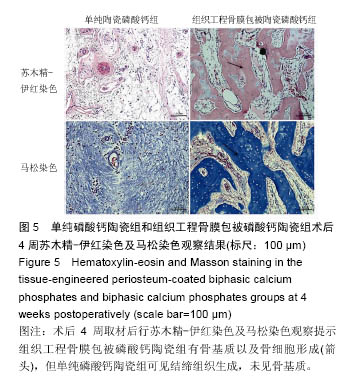
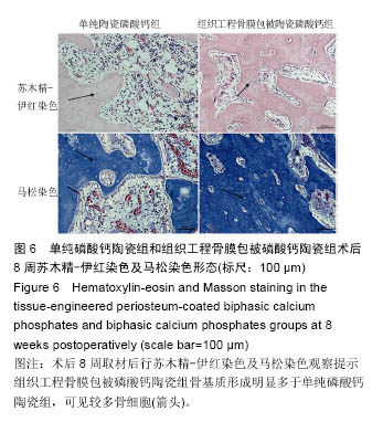
.jpg)
.jpg)