中国组织工程研究 ›› 2021, Vol. 25 ›› Issue (2): 211-215.doi: 10.3969/j.issn.2095-4344.2971

• 骨组织构建 bone tissue construction • 上一篇    下一篇

山茱萸新苷Ⅰ干预膝骨关节炎模型大鼠抑制白细胞介素6介导的炎性反应

李虎业1,窦增花1,孔德元1,蔡金莲1,李泽清2   

  1. 1 青海省第四人民医院药剂科,青海省西宁市   810000;2 青海大学附属医院,青海省西宁市   810000 
  • 收稿日期:2020-01-04 修回日期:2020-01-16 接受日期:2020-03-11 出版日期:2021-01-18 发布日期:2020-11-21
  • 通讯作者: 窦增花,副主任药师,青海省第四人民医院药剂科,青海省西宁市 810000
  • 作者简介:李虎业,男,1980年生,青海省人,汉族,2011年青海民族大学毕业,硕士,主管药师。
  • 基金资助:
    青海省基础研究计划项目(2018-0302-ZJC-0042)

Cornuside I inhibits interleukin-6-mediated inflammation in knee osteoarthritis rats

Li Huye1, Dou Zenghua1, Kong Deyuan1, Cai Jinlian1, Li Zeqing2    

  1. 1Pharmacy Department of the Fourth People’s Hospital of Qinghai Province, Xining 810000, Qinghai Province, China; 2Affiliated Hospital of Qinghai University, Xining 810000, Qinghai Province, China
  • Received:2020-01-04 Revised:2020-01-16 Accepted:2020-03-11 Online:2021-01-18 Published:2020-11-21
  • Contact: Dou Zenghua, Associate chief pharmacist, Pharmacy Department of the Fourth People’s Hospital of Qinghai Province, Xining 810000, Qinghai Province, China
  • About author:Li Huye, Master, Pharmacist-in-charge, Pharmacy Department of the Fourth People’s Hospital of Qinghai Province, Xining 810000, Qinghai Province, China
  • Supported by:
    the Basic Research Project of Qinghai Province, No. 2018-0302-ZJC-0042

摘要:

文题释义:
膝骨关节炎:为关节软骨变性、增生引起的慢性退行性骨病,临床主要表现为负重关节疼痛、肿胀或畸形。
山茱萸新苷:为传统配伍中药山茱萸中主要的抗炎、抗氧化成分,同时具有调节骨细胞增殖、降血糖、抗肿瘤和调节免疫等多种药理学活性。

背景:研究表明山茱萸新苷Ⅰ可促进成骨和软骨细胞的增殖,但对于修复膝骨关节炎早期软骨损伤的作用和机制尚不明确。
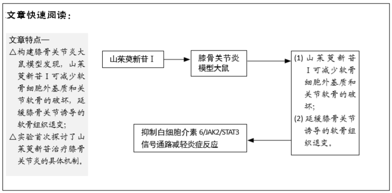
目的:观察山茱萸新苷Ⅰ对膝骨关节炎模型大鼠的作用及其对白细胞介素6/JAK2/STAT3信号通路的影响。
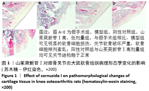
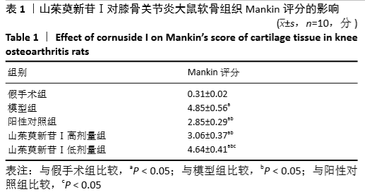
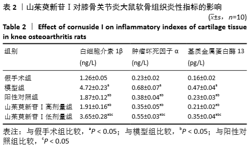
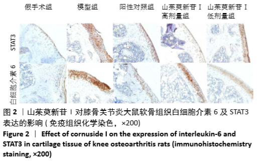
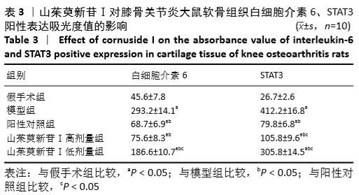
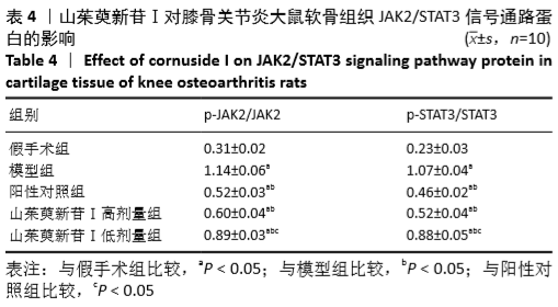
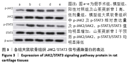
方法:将SD大鼠随机分为5组,除假手术组外,其他组均参照Hulth法制备膝骨关节炎大鼠模型,建模后山茱萸新苷Ⅰ低、高剂量组分别灌服山茱萸新苷Ⅰ1.25,5 g/kg,每日1次,阳性对照组则灌喂4 mg/kg的塞来昔布,假手术组与模型组则灌喂等量的生理盐水,每日1次,6周后于末次给药24 h后,苏木精-伊红染色观察大鼠关节软骨组织病理形态学变化,并依据Mankin’s法对大鼠软骨组织结构的退变程度进行评分;ELISA检测大鼠软骨组织中白细胞介素1β、肿瘤坏死因子α及基质金属蛋白酶13水平;免疫组织化学染色检测大鼠关节软骨组织中白细胞介素6、STAT3蛋白表达;Western blot法测定大鼠关节软骨组织中JAK2,p-JAK2,STAT3及p-STAT3蛋白相对表达量。
结果与结论:①模型组大鼠关节软骨破坏严重,可见明显的软骨细胞损伤,软骨细胞排列紊乱,山茱萸新苷Ⅰ低、高剂量组与阳性药对照组均可见关节结构趋于正常,软骨细胞分布趋于均匀,裂隙减小;②与模型组比较,山茱萸新苷Ⅰ低、高剂量组软骨组织Mankin评分、白细胞介素1β、肿瘤坏死因子α及基质金属蛋白酶13水平明显降低(P < 0.05),白细胞介素6和STAT3免疫反应均明显降低(P < 0.01),p-JAK2及p-STAT3蛋白相对表达量及p-JAK2/JAK2,p-STAT3/STAT3比值均明显降低(P < 0.05);(3)与阳性对照组相比,山茱萸新苷Ⅰ低剂量组的上述指标均增加(P < 0.05),山茱萸新苷Ⅰ高剂量组无显著差异;③结果证实,山茱萸新苷Ⅰ可延缓骨关节炎诱导的软骨组织退变,其机制可能与降低炎症因子表达,抑制白细胞介素6/JAK2/STAT3信号通路介导的炎症反应有关。
https://orcid.org/0000-0003-4883-3928 (李虎业) 

中国组织工程研究杂志出版内容重点:组织构建;骨细胞;软骨细胞;细胞培养;成纤维细胞;血管内皮细胞;骨质疏松;组织工程

关键词: 实验, 动物,  鼠,  因子,  蛋白,  通路,  膝,  骨关节炎

Abstract:

BACKGROUND: Studies have shown that cornuside I (Cor I) can promote the proliferation of osteoblasts and chondrocytes, but its role and mechanism in repairing early cartilage damage of knee osteoarthritis are not clear.

OBJECTIVE: To investigate the effect of Cor I on knee osteoarthritis rats and its effect on interleukin-6 (IL-6)/JAK2/STAT3 signaling pathway. 
METHODS: Fifty healthy male Sprague-Dawley rats were randomly divided into sham operation group, knee osteoarthritis model group (model group), Cor I high-dose group (Cor I-H), and cornuside I low-dose group (Cor I-L) and positive control group (celecoxib), with 10 rats in each group. Except the sham operation group, the knee osteoarthritis rat model was prepared according to the Hulth method. The Cor I-L and Cor I-H groups were administrated with Cor I 1.25 and 5 g/kg once a day for 6 weeks after modeling. The positive control group was fed with 4 mg/kg celecoxib, and the sham operation and model groups were fed with the same amount of normal saline once a day. The rats were sacrificed by cervical dislocation 24 hours after the last administration. Hematoxylin-eosin staining was used to observe the pathological and morphological changes of articular cartilage tissue in rats, and the degree of cartilage degeneration  was scored according to Mankin’s method. Interleukin-1β, tumor necrosis and matrix metalloproteinase-13 in rat cartilage tissue were detected by ELISA. Interleukin-6 and STAT3 protein expression in rat articular cartilage tissue were detected by immunohistochemistry. Relative expressions of JAK2, p-JAK2, STAT3, and p-STAT3 were detected by western blot. 
RESULTS AND CONCLUSION: The articular cartilage of rats was severely damaged in the model group, where obvious chondrocyte damage and disordered arrangement of chondrocytes were observed. The joint structure of the rats in the Cor I-H, Cor I-L and positive control groups tended to be normal, the chondrocytes were distributed uniformly, and the fissures were reduced. Compared with the model group, the relevant indicators in the Cor I-L and Cor I-H groups were significantly reduced, including the Mankin’s score of cartilage tissue, interleukin-1β, tumor necrosis factor α and matrix metalloproteinase-13 levels (P < 0.05), interleukin-6 and STAT3 immunoreactivities (P < 0.01) as well as the relative expression levels of p-JAK2 and p-STAT3 and ratios of p-JAK2/JAK2 and p-STAT3/STAT3 (P < 0.05). Compared with the positive control group, these indicators mentioned above were significantly increased in the Cor I-L group (P < 0.05); however, there was no significant difference between the positive control group and Cor I-H group. Results have confirmed that Cor I can delay knee osteoarthritis-induced cartilage tissue degeneration, and its mechanism may be related to reducing the were expression of inflammatory factors and inhibiting the inflammatory response mediated by interleukin-6/JAK2/STAT3 signaling pathway.

Key words: experiment, animal, rat, factor, protein, pathway, knee, osteoarthritis

中图分类号: